
|
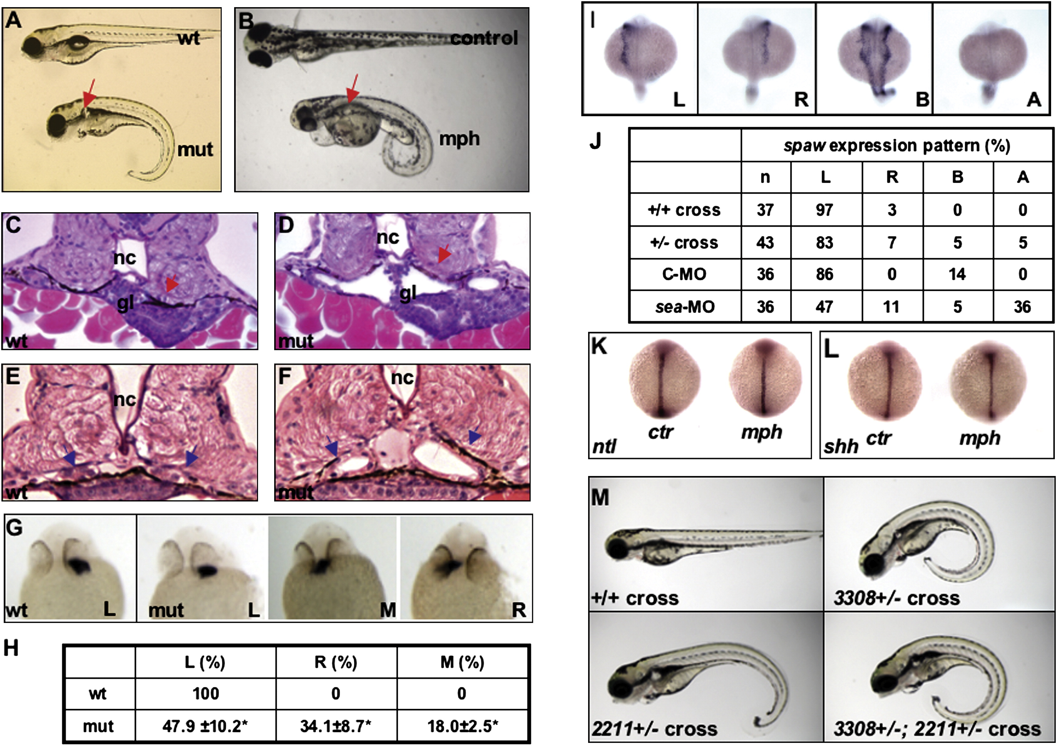
Fig. 1 Phenotypes Caused by Defective seahorse
(A) A seahorsehi3308 mutant on 5 dpf showing kidney cysts (arrow) and body curvature.
(B) A seahorse morphant on 3 dpf showing kidney cysts (arrow) and body curvature. An uninjected control sibling is also shown.
(C and D) Cross-section of the glomerular-tubular region of a wild-type and a seahorsehi3308 mutant embryo at 50 hpf. Arrows point to the glomerular and tubular region.
(E and F) Increased diameter of the pronephric duct (arrows) in a seahorsehi3308 mutant at 50 hpf, compared to a wild-type embryo.
(G–L) seahorse and left-right asymmetry. (G) In situ hybridization for cardiac myosin light chain (cmlc2) on embryos at 26 hpf showing the position of the heart tube. All are ventral views with head to the top. (H) Tabulation of heart position of embryos from three independent crosses. (I) In situ for spaw expression in lateral plate mesoderm on embryos at the 20-somite stage in dorsal views with head to the top. (J) Tabulation of spaw expression pattern. (K and L) The midline structure is not affected by sea-MO as shown by shh expression (K) and ntl expression (L) in embryos fixed at the 8-somite stage. Both in situ are shown in dorsal views with head to the top.
(M) seahorsehi3308 and ift172hi2211 double mutants show similar phenotypes as single mutants. All embryos are at 4 dpf, shown in side views with head to the left. Phenotypic embryos (body curvature and kidney cyst) are shown in all crosses except for the wild-type cross.
wt, wild-type; mut, mutant; mph, morphant; ctr, embryos injected with the control morpholino oligo; nc, notochord; gl, glomeruli; L, left side; M, middle; R, right side; A, absent; C-MO, embryos injected with a standard control oligo; sea-MO, embryos injected with seahorse morpholino; n, number of embryos analyzed.
Reprinted from Developmental Cell, 14(6), Kishimoto, N., Cao, Y., Park, A., and Sun, Z., Cystic kidney gene seahorse regulates cilia-mediated processes and Wnt pathways, 954-961, Copyright (2008) with permission from Elsevier. Full text @ Dev. Cell