
Image
|
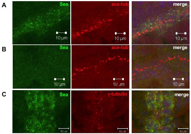
Figure Caption
Fig. S2
Details for Seahorse Subcellular Localization
(A-B) Split channel images of confocal projections of immuno-staining for Seahorse in the pronephric duct in embryos injected with a control morpholino oligo (A) or sea-MO (B) at the 18-somite stage. Seahorse signal is shown in green, while the cilia marker antiacetylated tubulin is shown in red. (C) Split channel images of confocal projections of immuno-staining of Seahorse in the pronephric duct in an embryo at the 18-somite stage. Seahorse signal is shown in green, while the centrosome/basal body marker anti-γ tubulin is shown in red.
Figure Data
Acknowledgments
This image is the copyrighted work of the attributed author or publisher, and
ZFIN has permission only to display this image to its users.
Additional permissions should be obtained from the applicable author or publisher of the image.
Reprinted from Developmental Cell, 14(6), Kishimoto, N., Cao, Y., Park, A., and Sun, Z., Cystic kidney gene seahorse regulates cilia-mediated processes and Wnt pathways, 954-961, Copyright (2008) with permission from Elsevier. Full text @ Dev. Cell