- Title
-
Genes involved in forebrain development in the zebrafish, Danio rerio
- Authors
- Heisenberg, C.P., Brand, M., Jiang, Y.J., Warga, R.M., Beuchle, D., van Eeden, F.J., Furutani-Seiki, M., Granato, M., Haffter, P., Hammerschmidt, M., Kane, D.A., Kelsh, R.N., Mullins, M.C., Odenthal, J., and Nüsslein-Volhard, C.
- Source
- Full text @ Development
|
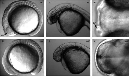
Phenotypes of live wild type (A-C) and mbl (D-F) embryos. (A,D) Lateral view of 14-hour embryos showing the absence of optic vesicles in mbl. (B,E) Lateral view of 24-hour embryos showing the absence of eyes in mbl. (C,F) Dorsal view of 96-hour embryos showing the absence of olfactory placodes in mbl. OV, optic vesicles; OP, olfactory placodes. Anterior to the left. PHENOTYPE:
|
|
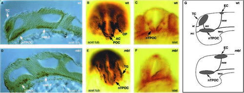
Whole-mount antibody stainings of 24-hour embryos for acetylated tubulin and islet proteins (anti-pan-islet antibody) visualizing primary neurons and their axons in wild-type (A-C) and mbl (D-F) embryos. (A,D) Sagittal sections through the head region stained for acetylated tubulin showing the absence of the telencephalic neuronal clusters in mbl embryos (anterior to the left). (B,E) Frontal view of the head of embryos stained for acetylated tubulin showing the absence of olfactory placodes, and anterior postoptic commissures and the expansion of the trigeminal ganglia in mbl embryos. (C,F) Frontal views of the head (optical section) of embryos stained for islet proteins showing the absence of islet-positive cells within the nTPOC in mbl. (G) Schematic drawings of lateral views of the head summarizing the results from A-F (anterior to the left). TC, telencephalic cluster; nTPOC, nucleus of the tract of the postoptic commissure; nMLF, nucleus of the medial longitudinal fisciculus; EC, epiphysial cluster; AC, anterior commissure, POC, postoptic commissure; SOT, supraoptic tract; TPOC, tract of the postoptic commissure; TG, trigeminal ganglion; OP, olfactory placodes; DVDT, dorsal ventral diencephalic tract. |
|
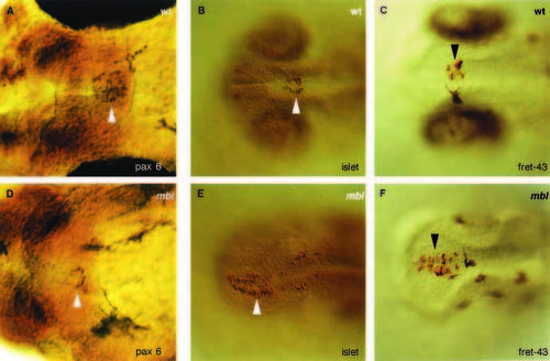
Whole-mount antibody stainings of embryos for pax6 (30 hour), islet proteins (antipan- islet antibody; 24 hour), and fret43 (24 hour) visualizing the anterior pituitary and epiphysis in wild-type (A-C) and mbl (D-F) embryos. (A,D) Ventral view of the head of embryos stained for pax6 showing a reduction in the number of pax6-positive neurons within the anterior pituitary (arrowhead) of mbl embryos. (B,C,E,F) Dorsal view of the head of embryos stained for islet proteins (B,E) and fret43 (C,F) showing an increase in the number of islet/fret43-positive cells within the epiphysis (arrowhead) of mbl. Anterior to the left. |
|
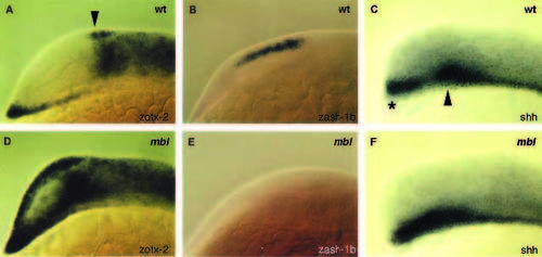
Whole-mount in situ labeling of embryos visualizing the forebrain expression domains of zotx-2 (14 hour), zash-1b (12 hour), and shh (14 hour) of wild-type (A-C) and mbl (D-F) embryos. (A,D) Lateral view of the head showing an expansion of the zotx-2 expression domain anteriorly in mbl (arrowhead points to the epiphysis anlage in wild type). (B,E) Lateral view of the head showing the absence of the telencephalic expression domain of zash-1b in mbl. (C,F) Lateral view of the head showing an expansion of the hypothalamic (asterisk) and/or diencephalic expression domain (arrowhead) of shh in mbl. Anterior to the left. EXPRESSION / LABELING:
PHENOTYPE:
|
|
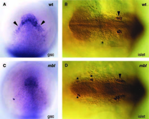
Whole-mount antibody and in situ stainings of embryos for islet proteins (anti-pan-islet-antibody) (12 hour) and gsc (10 hour) visualizing the trigeminal placodes (islet) and prechordal plate (gsc) of wild type (A,B) and mbl (C,D) embryos. (A,C) Dorsal view of the head showing a reduction of gsc expression overlying the anteriorlateral edge of the prechordal plate (arrowheads) in mbl (anterior up). (B,D) Dorsal view of the head showing ectopic clusters of isletpositive cells (stars) anterior to the trigeminal placodes (arrowhead) in mbl (anterior to the left). EXPRESSION / LABELING:
PHENOTYPE:
|
|
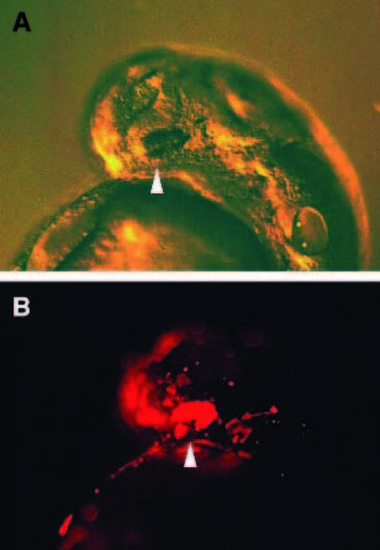
Transplantation of rhodamine-dextran labeled wild-type cells into mbl mutants. (A) Phenotype of a live, rescued, 30-hour mbl embryo showing small eyes (arrowhead). (B) UV light image of the same embryo showing that the rescued eye is exclusively formed of labeled (transplanted wild-type) cells (arrowhead). Anterior to the left. |
|
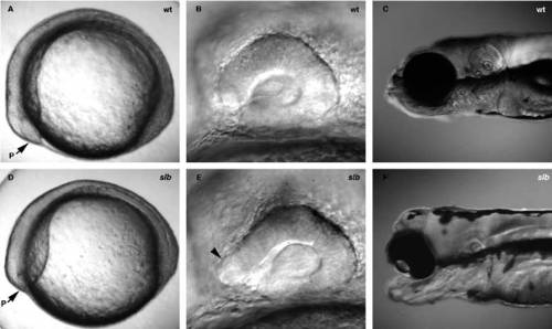
Phenotypes of live wildtype (A-C) and slb embryos (D-F). (A,D) Lateral views of 11-hour embryos showing a reduced body length and a smaller polster in slb. (B,E) Lateral views of the head of 24-hour embryos showing a fusion of the eyes in slb (arrowhead points to the region of the optic stalks in slb). (C,F) Lateral views of the head of 120-hour embryos showing a fusion of the eyes and deformation of the jaw in slb embryos. P, polster. Anterior to the left. PHENOTYPE:
|
|
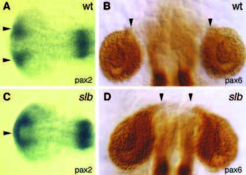
Whole-mount antibody stainings for pax6 and in situ stainings forpax6 of 20-hour embryos visualizing the optic stalks and retinae of wild-type (A,B) and slb (C,D) embryos. (A,C) Dorsal views of the head stained for pax2 showing a fusion of the optic stalk region in slb (anterior to the left). (B,D) Dorsal views of the head stained for pax6 showing separated retinae in slb. Arrowheads indicate the region of the optic stalks (anterior up). EXPRESSION / LABELING:
PHENOTYPE:
|
|
Whole-mount antibody and in situ stainings of 10-hour embryos visualizing the expression of ntl and myoD in wildtype (A,B) and slb (A,C) embryos. (A) Dorsal view showing that the notochord anlage stained for ntl is broadened in slb embryos (anterior to the right). (B,C) Dorsal view of double labeled embryos (antibody and in situ) showing that the notochord anlage (ntl, brown colour) is broadened in slb. At the same time myoD expression (blue colour) in the presomitic mesoderm of slb embryos looks wild type (anterior up). |
|
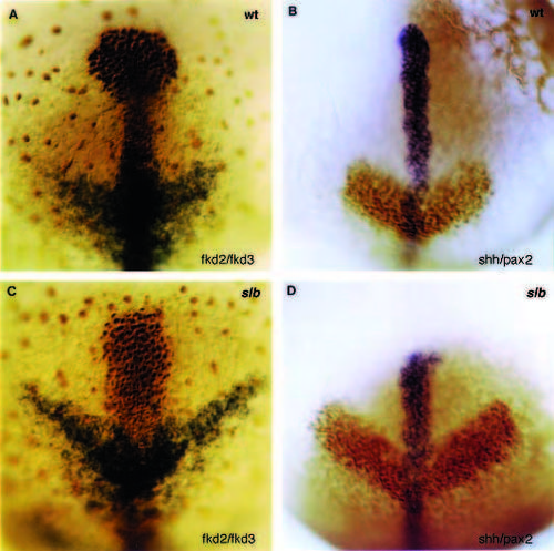
Whole-mount antibody and in situ stainings of 10-hour embryos visualizing the expression domains of fkd2, fkd3, pax2 and shh in wild-type (A,B) and slb (C,D) embryos. (A,C) Dorsal views of the head of double labeled (antibody and in situ) embryos showing an altered shape of the prechordal plate stained for fkd2 (brown colour) and a broadened neuroectodermal expression domain of fkd3 at the level of the diencephalon (blue colour) in slb. (B,D) Dorsal views of the head of double labeled (antibody and in situ) embryos showing a shortened expression domain of shh in the anterior-ventral neuroectoderm (blue colour) and broadened neuroectodermal expression domain of pax2 (brown colour) at the level of the midhindbrain- boundary anlage in slb. Anterior up. |
|
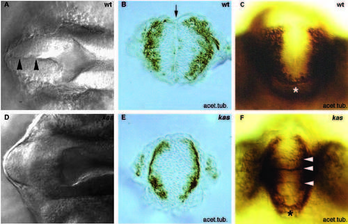
Phenotype of live embryos (A,D) and stainings for acetylated tubulin (B,C,E,F) in 24-hour wild type (A-C) and kas (D-F) embryos. (A,D) Dorsal views of the head showing the absence of the dorsal midline in the anterior forebrain (black arrowheads in wild type) in kas (anterior to the left). (B,E) Frontal sections of the head at the level of the anterior telencephalon of embryos stained for acetylated tubulin showing a disorganization of cells at the persumptive midline (arrow in wild type) between these clusters in kas. (C,F) Frontal views of the head of embryos stained for acetylated tubulin showing that the anterior commissure (white arrowheads) is defasciculated in kas. Star indicates the position of the anterior commissure in wild type. PHENOTYPE:
|
|
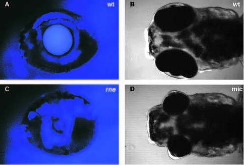
Phenotypes of live120-hour wild type (A) and rne (C) embryos. (A,C) Lateral views of the eye (UV images) showing degenerating lenses in rne embryos. Phenotypes of live120-hour wild type (B) and mic (D) embryos. (B,D) Dorsal view of the head showing smaller eyes in mic embryos. Anterior to the left. PHENOTYPE:
|

