
Image
|
Figure Caption
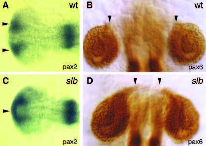
Fig. 8
Whole-mount antibody stainings for pax6 and in situ stainings forpax6 of 20-hour embryos visualizing the optic stalks and retinae of wild-type (A,B) and slb (C,D) embryos. (A,C) Dorsal views of the head stained for pax2 showing a fusion of the optic stalk region in slb (anterior to the left). (B,D) Dorsal views of the head stained for pax6 showing separated retinae in slb. Arrowheads indicate the region of the optic stalks (anterior up).
Figure Data
Acknowledgments
This image is the copyrighted work of the attributed author or publisher, and
ZFIN has permission only to display this image to its users.
Additional permissions should be obtained from the applicable author or publisher of the image.
Full text @ Development