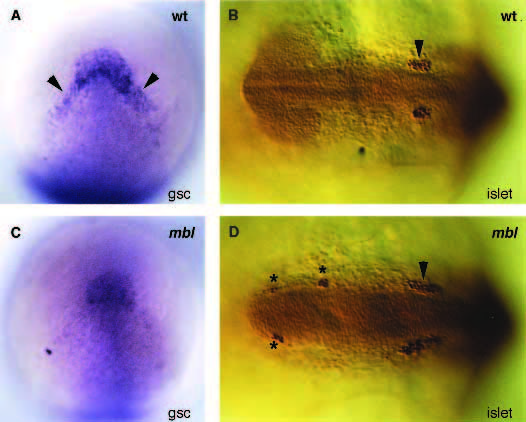
Image


                        Figure Caption


Fig. 5
Whole-mount antibody and in situ stainings of embryos for islet proteins (anti-pan-islet-antibody) (12 hour) and gsc (10 hour) visualizing the trigeminal placodes (islet) and prechordal plate (gsc) of wild type (A,B) and mbl (C,D) embryos. (A,C) Dorsal view of the head showing a reduction of gsc expression overlying the anteriorlateral edge of the prechordal plate (arrowheads) in mbl (anterior up). (B,D) Dorsal view of the head showing ectopic clusters of isletpositive cells (stars) anterior to the trigeminal placodes (arrowhead) in mbl (anterior to the left).
                
                    
                        Figure Data


                        Acknowledgments


This image is the copyrighted work of the attributed author or publisher, and
ZFIN has permission only to display this image to its users.
Additional permissions should be obtained from the applicable author or publisher of the image.
      Full text @ Development