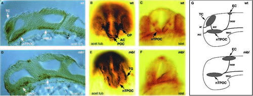
Whole-mount antibody stainings of 24-hour embryos for acetylated tubulin and islet proteins (anti-pan-islet antibody) visualizing primary neurons and their axons in wild-type (A-C) and mbl (D-F) embryos. (A,D) Sagittal sections through the head region stained for acetylated tubulin showing the absence of the telencephalic neuronal clusters in mbl embryos (anterior to the left). (B,E) Frontal view of the head of embryos stained for acetylated tubulin showing the absence of olfactory placodes, and anterior postoptic commissures and the expansion of the trigeminal ganglia in mbl embryos. (C,F) Frontal views of the head (optical section) of embryos stained for islet proteins showing the absence of islet-positive cells within the nTPOC in mbl. (G) Schematic drawings of lateral views of the head summarizing the results from A-F (anterior to the left). TC, telencephalic cluster; nTPOC, nucleus of the tract of the postoptic commissure; nMLF, nucleus of the medial longitudinal fisciculus; EC, epiphysial cluster; AC, anterior commissure, POC, postoptic commissure; SOT, supraoptic tract; TPOC, tract of the postoptic commissure; TG, trigeminal ganglion; OP, olfactory placodes; DVDT, dorsal ventral diencephalic tract.
|
