
|
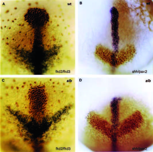
Fig. 10
Whole-mount antibody and in situ stainings of 10-hour embryos visualizing the expression domains of fkd2, fkd3, pax2 and shh in wild-type (A,B) and slb (C,D) embryos. (A,C) Dorsal views of the head of double labeled (antibody and in situ) embryos showing an altered shape of the prechordal plate stained for fkd2 (brown colour) and a broadened neuroectodermal expression domain of fkd3 at the level of the diencephalon (blue colour) in slb. (B,D) Dorsal views of the head of double labeled (antibody and in situ) embryos showing a shortened expression domain of shh in the anterior-ventral neuroectoderm (blue colour) and broadened neuroectodermal expression domain of pax2 (brown colour) at the level of the midhindbrain- boundary anlage in slb. Anterior up.