FIGURE SUMMARY
Title

Regulation of the apical extension morphogenesis tunes the mechanosensory response of microvilliated neurons

Authors
Desban, L., Prendergast, A., Roussel, J., Rosello, M., Geny, D., Wyart, C., Bardet, P.L.
Source
Full text @ PLoS Biol.

CSF-cNs go through 3 critical steps to form their apical extension.

Z-projections from time-lapse acquisitions (top panels; lateral views of ventral CSF-cNs with rostral to the left) and schematics (bottom panels) showing the 3 stages CSF-cNs go through during the formation of their AE. The CSF-cN soma becomes round and short actin-based protrusions (Stage 2) arise from the ring of actin (Stage 1, arrowhead) concomitantly with a subapical constriction (Stages 2 and 3; chevrons). Gradually, protrusions lengthen to form the AE (Stage 3; arrow) characteristic of differentiated CSF-cNs. Data collected over 13 time-lapse sessions for 15 ventral and 6 dorsolateral cells. The number of cells imaged transitioning from one stage to the next is indicated in the bottom legend. Scale bar, 10 μm. AE, apical extension; CSF-cN, cerebrospinal fluid-contacting neuron. hpf, hours post fertilization.

The ring of actin colocalizes with the CSF-cN apical junctional complexes.

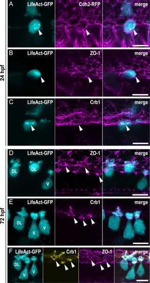
(A–C) Z-projections from lateral views of the spinal cord in 24-hpf embryos showing the colocalization of the ring of actin (LifeAct; arrowheads) with different markers of the AJCs: (A) Cdh2 for adherens junctions, (B) ZO-1 for tight junctions, and (C) Crb1 for the apical domain. (A) Double immunostaining for GFP and RFP in triple transgenic Tg(pkd2l1:Gal4,UAS:LifeAct-GFP;cryaa:V,cdh2:cdh2-RFP) embryos. (B, C) Double immunostaining for GFP and (B) ZO-1, or (C) Crb1 in Tg(pkd2l1:Gal4,UAS:LifeAct-GFP;cryaa:V) embryos. (D–F) Z-projections from lateral views of 72-hpf Tg(pkd2l1:Gal4,UAS:LifeAct-GFP;cryaa:V) larvae immunostained for GFP and (D) ZO-1, (E) Crb1, or (F) ZO-1 and Crb1. The markers ZO-1 and Crb1 are retained at the AJCs (arrowheads) in both V and DL CSF-cNs after differentiation. In DL cells, Crb1 expands to the apical extension. Scale bars, 10 μm. AJC, apical junctional complexes; Cdh2, Cadherin 2; Crb1, Crumbs 1; CSF-cN, cerebrospinal fluid-contacting neurons; DL, dorso-lateral; GFP, green fluorescent protein; hpf, hours post fertilization; RFP, red fluorescent protein; V, ventral; ZO-1, zonula-occludens-1.

Crb1 participates in the proper development of the CSF-cN apical extension.

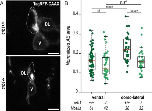
(A) Z-projections from transversal sections showing V and DL TagRFP-CAAX-expressing CSF-cNs in 72-hpf larvae illustrating the smaller AE in crb1−/− (bottom panel) compared with wild-type siblings (top panel). The central canal is outlined (dotted lines) according to ZO-1 staining. Scale bars, 5 μm. (B) Quantification of the normalized area covered by CSF-cN AEs at 72 hpf in V and DL cells in crb1−/− (light green; N = 3 fish) compared with wild-type siblings (dark green; N = 4 fish). In both CSF-cN subtypes, the AE was significantly smaller in mutant larvae compared with wild-type (p1 = 0.0477, p2 = 9.9019 × 10⁻⁵, p3 = 2.6381 × 10⁻⁵, p4 = 0.1876). Underlying data can be found in S1 Data. AE, apical extension; Crb1, Crumbs 1; CSF-cN, cerebrospinal fluid-contacting neuron; DL, dorsolateral; hpf, hours post fertilization; n.s., not significant; V, ventral; ZO-1, zonula-occludens-1.

CSF-cNs express a set of known morphogenetic factors.

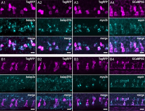
Expression of candidate factors known to be involved in the formation of actin-based protrusions—baiap2a, baiap2l1b, myo3b, and espin—assessed by FISH at 24 hpf (A1–4) and 72 hpf (B1–4). Expression in CSF-cNs was validated by combining FISH to IHC for RFP or GFP in Tg(pkd2l1:TagRFP) (A1–3, B1–3) or Tg(pkd2l1:GCaMP5G) (A4, B4) transgenic animals, respectively. In 24-hpf embryos, baipa2a (A1), baiap2l1b (A2), myo3b (A3), and espin (A4) were enriched in CSF-cNs. In 72-hpf larvae, expression of baiap2a (B1) and baiap2l1b (B2) was not clearly detected, whereas myo3b (B3) and espin (B4) remained strongly expressed in CSF-cNs. Scale bars, 10 μm. CSF-cN, cerebrospinal fluid-contacting neuron; FISH, fluorescent in situ hybridization; GFP, green fluorescent protein; hpf, hours post fertilization; IHC, immunohistochemistry; RFP, red fluorescent protein.

In the absence of Espin actin-bundling activity, CSF-cNs form shorter apical extensions.

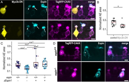
(A) Z-projections of whole-mounted spinal cords at 72 hpf showing mosaic expression of Myo3b-DN under the control of the pkd2l1 promoter. Immunostaining reveals Espin (cyan) in TagRFP-CAAX-positive CSF-cNs (magenta) expressing Myo3b-DN (yellow, arrowheads) or not (arrows). In ventral CSF-cNs expressing Myo3b-DN, Espin staining is reduced (observed in 9 cells out of 9), and the AE appears smaller compared with wild-type cells. (B) Quantification of the normalized area covered by the AE of ventral cells expressing Myo3b-DN (N = 9 cells) compared with nonexpressing neighboring cells (control; N = 10 cells). Cells expressing Myo3b-DN form a significantly smaller AE (N = 5 fish; p = 0.025). (C) The same quantification was performed in espin−/− ventral cells at 72 hpf (N = 30 cells in 5 fish) compared with wild-type cells (N = 16 cells in 5 fish). Mutant cells display significantly smaller AEs (p1 = 8.4010 × 10⁻⁶). In wild-type ventral cells, the overexpression of the zEspin1 was sometimes associated with abnormally long microvilli (observed in 2 cells out of 9; red chevrons; p2 = 0.2123). In espin−/ cells, the mutant phenotype was rescued by zEspin1 (N = 15 cells; p3 = 0.3029 and p4 = 3.8556 × 10⁻⁶). (D) Z-projections of whole-mounted spinal cords at 72 hpf showing mosaic expression of zEspin1 under the control of the pkd2l1 promoter. Immunostaining for Espin (cyan) in TagRFP-CAAX-positive ventral CSF-cNs (magenta) reveals the loss of Espin immunoreactivity in espin−/− larvae, which is retrieved in mutant cells expressing zEspin1 (nuclear RFP; magenta; stars). Scale bars, 5 μm. Underlying data can be found in S1 Data. AE, apical extension; CSF-cN, cerebrospinal fluid-contacting neuron; DN, dominant-negative; hpf, hours post fertilization; Myo3b, myosin 3b;n.s., not significant; RFP, red fluorescent protein; zEspin1, zebrafish Espin isoform 1.

CSF-cNs with shorter apical extensions exhibit reduced mechanosensory response.

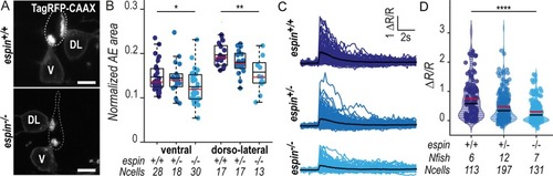
(A) Z-projections from transversal sections of spinal cords with V and DL TagRFP-CAAX-positive CSF-cNs at 72 hpf illustrating the smaller AEs in espin−/− (bottom panel) compared with wild-type siblings (top panel). The central canal was outlined (dotted lines) according to ZO-1 staining. Scale bars, 5 μm. (B) Quantification of the normalized area covered by the CSF-cN AE at 72 hpf in V and DL cells in espin−/− (light blue, N = 2 fish), espin+/− (blue, N = 2 fish), and espin+/+ (dark blue, N = 3 fish) siblings (1 representative experiment out of 2). In both CSF-cN subtypes, the AE size gradually decreases when cells miss 1 (espin+/−) or 2 (espin−/−) copies of the wild-type allele (pV = 0.0112, df = 74, and t = 2.3311 in ventral cells; pDL = 0.0017, df = 45, and t = 3.0935 in DL cells). (C) Overlay of calcium transients in ipsilateral DL CSF-cNs in response to passive tail bending induced by a glass probe in paralyzed control wild-type larvae versus espin+/− and espin−/− siblings at 5 days (120 hpf). The average across cells is shown in black (pulled data from 4 experiments). (D) The amplitude of CSF-cN calcium transients shown in (C) is represented as the ratio of peak fluorescence over baseline (ΔR/R) and is gradually reduced in espin+/− and espin−/− compared with wild-type siblings in a wild-type allele number-dependent manner (p = 1.1102 × 10⁻⁶, df = 1309, and t = 8.462). Underlying data can be found in S1 Data. AE, apical extension; CSF-cN, cerebrospinal fluid-contacting neuron; df, degrees of freedom; DL, dorsolateral; hpf, hours post fertilization; V, ventral; ZO-1, zonula-occludens-1.

Acknowledgments
This image is the copyrighted work of the attributed author or publisher, and ZFIN has permission only to display this image to its users. Additional permissions should be obtained from the applicable author or publisher of the image. Full text @ PLoS Biol.