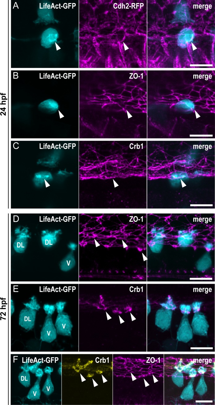
IMAGE

Fig 2

ID
ZDB-IMAGE-190723-1224
Genes
Antibodies
Source
Figures for Desban et al., 2019
Image
Figure Caption

Fig 2 The ring of actin colocalizes with the CSF-cN apical junctional complexes.

(A–C) Z-projections from lateral views of the spinal cord in 24-hpf embryos showing the colocalization of the ring of actin (LifeAct; arrowheads) with different markers of the AJCs: (A) Cdh2 for adherens junctions, (B) ZO-1 for tight junctions, and (C) Crb1 for the apical domain. (A) Double immunostaining for GFP and RFP in triple transgenic Tg(pkd2l1:Gal4,UAS:LifeAct-GFP;cryaa:V,cdh2:cdh2-RFP) embryos. (B, C) Double immunostaining for GFP and (B) ZO-1, or (C) Crb1 in Tg(pkd2l1:Gal4,UAS:LifeAct-GFP;cryaa:V) embryos. (D–F) Z-projections from lateral views of 72-hpf Tg(pkd2l1:Gal4,UAS:LifeAct-GFP;cryaa:V) larvae immunostained for GFP and (D) ZO-1, (E) Crb1, or (F) ZO-1 and Crb1. The markers ZO-1 and Crb1 are retained at the AJCs (arrowheads) in both V and DL CSF-cNs after differentiation. In DL cells, Crb1 expands to the apical extension. Scale bars, 10 μm. AJC, apical junctional complexes; Cdh2, Cadherin 2; Crb1, Crumbs 1; CSF-cN, cerebrospinal fluid-contacting neurons; DL, dorso-lateral; GFP, green fluorescent protein; hpf, hours post fertilization; RFP, red fluorescent protein; V, ventral; ZO-1, zonula-occludens-1.

Figure Data
Acknowledgments
This image is the copyrighted work of the attributed author or publisher, and ZFIN has permission only to display this image to its users. Additional permissions should be obtained from the applicable author or publisher of the image. Full text @ PLoS Biol.