
|
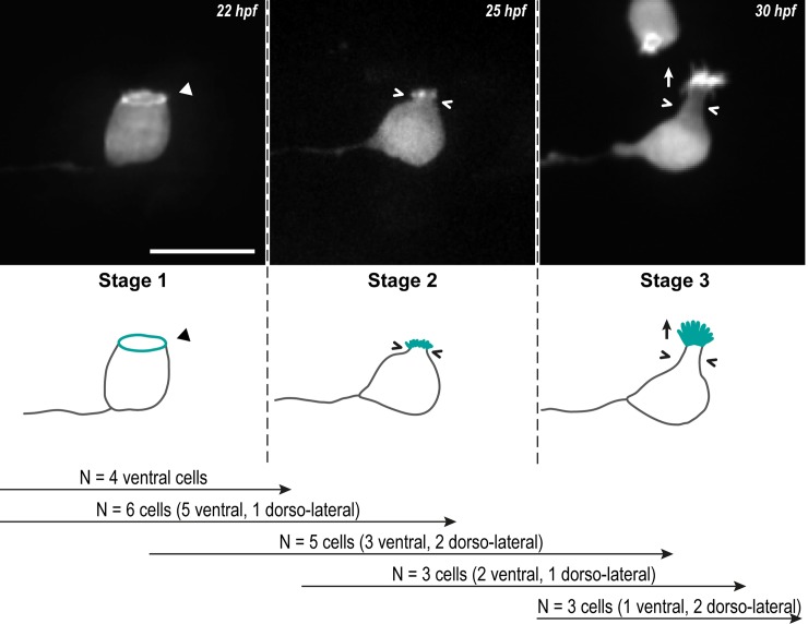
Fig 1
Z-projections from time-lapse acquisitions (top panels; lateral views of ventral CSF-cNs with rostral to the left) and schematics (bottom panels) showing the 3 stages CSF-cNs go through during the formation of their AE. The CSF-cN soma becomes round and short actin-based protrusions (Stage 2) arise from the ring of actin (Stage 1, arrowhead) concomitantly with a subapical constriction (Stages 2 and 3; chevrons). Gradually, protrusions lengthen to form the AE (Stage 3; arrow) characteristic of differentiated CSF-cNs. Data collected over 13 time-lapse sessions for 15 ventral and 6 dorsolateral cells. The number of cells imaged transitioning from one stage to the next is indicated in the bottom legend. Scale bar, 10 μm. AE, apical extension; CSF-cN, cerebrospinal fluid-contacting neuron. hpf, hours post fertilization.