IMAGE

Fig 6

ID
ZDB-IMAGE-190723-1230
Source
Figures for Desban et al., 2019
Image
Figure Caption

Fig 6 CSF-cNs with shorter apical extensions exhibit reduced mechanosensory response.

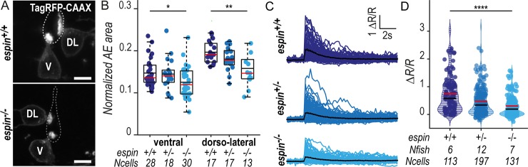
(A) Z-projections from transversal sections of spinal cords with V and DL TagRFP-CAAX-positive CSF-cNs at 72 hpf illustrating the smaller AEs in espin−/− (bottom panel) compared with wild-type siblings (top panel). The central canal was outlined (dotted lines) according to ZO-1 staining. Scale bars, 5 μm. (B) Quantification of the normalized area covered by the CSF-cN AE at 72 hpf in V and DL cells in espin−/− (light blue, N = 2 fish), espin+/− (blue, N = 2 fish), and espin+/+ (dark blue, N = 3 fish) siblings (1 representative experiment out of 2). In both CSF-cN subtypes, the AE size gradually decreases when cells miss 1 (espin+/−) or 2 (espin−/−) copies of the wild-type allele (pV = 0.0112, df = 74, and t = 2.3311 in ventral cells; pDL = 0.0017, df = 45, and t = 3.0935 in DL cells). (C) Overlay of calcium transients in ipsilateral DL CSF-cNs in response to passive tail bending induced by a glass probe in paralyzed control wild-type larvae versus espin+/− and espin−/− siblings at 5 days (120 hpf). The average across cells is shown in black (pulled data from 4 experiments). (D) The amplitude of CSF-cN calcium transients shown in (C) is represented as the ratio of peak fluorescence over baseline (ΔR/R) and is gradually reduced in espin+/− and espin−/− compared with wild-type siblings in a wild-type allele number-dependent manner (p = 1.1102 × 10⁻⁶, df = 1309, and t = 8.462). Underlying data can be found in S1 Data. AE, apical extension; CSF-cN, cerebrospinal fluid-contacting neuron; df, degrees of freedom; DL, dorsolateral; hpf, hours post fertilization; V, ventral; ZO-1, zonula-occludens-1.

Figure Data
Acknowledgments
This image is the copyrighted work of the attributed author or publisher, and ZFIN has permission only to display this image to its users. Additional permissions should be obtained from the applicable author or publisher of the image. Full text @ PLoS Biol.