|
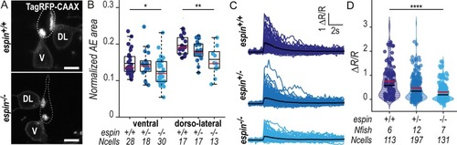
CSF-cNs with shorter apical extensions exhibit reduced mechanosensory response.(A) Z-projections from transversal sections of spinal cords with V and DL TagRFP-CAAX-positive CSF-cNs at 72 hpf illustrating the smaller AEs in espin−/− (bottom panel) compared with wild-type siblings (top panel). The central canal was outlined (dotted lines) according to ZO-1 staining. Scale bars, 5 μm. (B) Quantification of the normalized area covered by the CSF-cN AE at 72 hpf in V and DL cells in espin−/− (light blue, N = 2 fish), espin+/− (blue, N = 2 fish), and espin+/+ (dark blue, N = 3 fish) siblings (1 representative experiment out of 2). In both CSF-cN subtypes, the AE size gradually decreases when cells miss 1 (espin+/−) or 2 (espin−/−) copies of the wild-type allele (pV = 0.0112, df = 74, and t = 2.3311 in ventral cells; pDL = 0.0017, df = 45, and t = 3.0935 in DL cells). (C) Overlay of calcium transients in ipsilateral DL CSF-cNs in response to passive tail bending induced by a glass probe in paralyzed control wild-type larvae versus espin+/− and espin−/− siblings at 5 days (120 hpf). The average across cells is shown in black (pulled data from 4 experiments). (D) The amplitude of CSF-cN calcium transients shown in (C) is represented as the ratio of peak fluorescence over baseline (ΔR/R) and is gradually reduced in espin+/− and espin−/− compared with wild-type siblings in a wild-type allele number-dependent manner (p = 1.1102 × 10⁻⁶, df = 1309, and t = 8.462). Underlying data can be found in S1 Data. AE, apical extension; CSF-cN, cerebrospinal fluid-contacting neuron; df, degrees of freedom; DL, dorsolateral; hpf, hours post fertilization; V, ventral; ZO-1, zonula-occludens-1.
|