IMAGE

Fig 3

ID
ZDB-IMAGE-190723-1227
Source
Figures for Desban et al., 2019
Image
Figure Caption

Fig 3 Crb1 participates in the proper development of the CSF-cN apical extension.

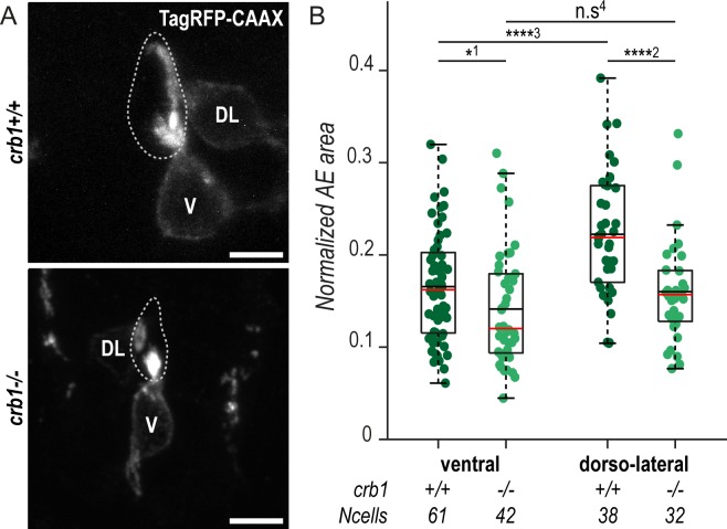
(A) Z-projections from transversal sections showing V and DL TagRFP-CAAX-expressing CSF-cNs in 72-hpf larvae illustrating the smaller AE in crb1−/− (bottom panel) compared with wild-type siblings (top panel). The central canal is outlined (dotted lines) according to ZO-1 staining. Scale bars, 5 μm. (B) Quantification of the normalized area covered by CSF-cN AEs at 72 hpf in V and DL cells in crb1−/− (light green; N = 3 fish) compared with wild-type siblings (dark green; N = 4 fish). In both CSF-cN subtypes, the AE was significantly smaller in mutant larvae compared with wild-type (p1 = 0.0477, p2 = 9.9019 × 10⁻⁵, p3 = 2.6381 × 10⁻⁵, p4 = 0.1876). Underlying data can be found in S1 Data. AE, apical extension; Crb1, Crumbs 1; CSF-cN, cerebrospinal fluid-contacting neuron; DL, dorsolateral; hpf, hours post fertilization; n.s., not significant; V, ventral; ZO-1, zonula-occludens-1.

Figure Data
Acknowledgments
This image is the copyrighted work of the attributed author or publisher, and ZFIN has permission only to display this image to its users. Additional permissions should be obtained from the applicable author or publisher of the image. Full text @ PLoS Biol.