IMAGE

Fig 5

ID
ZDB-IMAGE-190723-1229
Source
Figures for Desban et al., 2019
Image
Figure Caption

Fig 5 In the absence of Espin actin-bundling activity, CSF-cNs form shorter apical extensions.

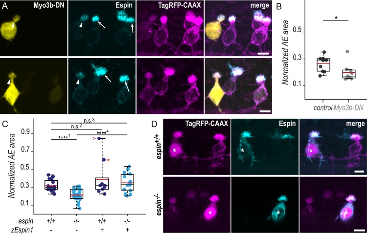
(A) Z-projections of whole-mounted spinal cords at 72 hpf showing mosaic expression of Myo3b-DN under the control of the pkd2l1 promoter. Immunostaining reveals Espin (cyan) in TagRFP-CAAX-positive CSF-cNs (magenta) expressing Myo3b-DN (yellow, arrowheads) or not (arrows). In ventral CSF-cNs expressing Myo3b-DN, Espin staining is reduced (observed in 9 cells out of 9), and the AE appears smaller compared with wild-type cells. (B) Quantification of the normalized area covered by the AE of ventral cells expressing Myo3b-DN (N = 9 cells) compared with nonexpressing neighboring cells (control; N = 10 cells). Cells expressing Myo3b-DN form a significantly smaller AE (N = 5 fish; p = 0.025). (C) The same quantification was performed in espin−/− ventral cells at 72 hpf (N = 30 cells in 5 fish) compared with wild-type cells (N = 16 cells in 5 fish). Mutant cells display significantly smaller AEs (p1 = 8.4010 × 10⁻⁶). In wild-type ventral cells, the overexpression of the zEspin1 was sometimes associated with abnormally long microvilli (observed in 2 cells out of 9; red chevrons; p2 = 0.2123). In espin−/ cells, the mutant phenotype was rescued by zEspin1 (N = 15 cells; p3 = 0.3029 and p4 = 3.8556 × 10⁻⁶). (D) Z-projections of whole-mounted spinal cords at 72 hpf showing mosaic expression of zEspin1 under the control of the pkd2l1 promoter. Immunostaining for Espin (cyan) in TagRFP-CAAX-positive ventral CSF-cNs (magenta) reveals the loss of Espin immunoreactivity in espin−/− larvae, which is retrieved in mutant cells expressing zEspin1 (nuclear RFP; magenta; stars). Scale bars, 5 μm. Underlying data can be found in S1 Data. AE, apical extension; CSF-cN, cerebrospinal fluid-contacting neuron; DN, dominant-negative; hpf, hours post fertilization; Myo3b, myosin 3b;n.s., not significant; RFP, red fluorescent protein; zEspin1, zebrafish Espin isoform 1.

Figure Data
Acknowledgments
This image is the copyrighted work of the attributed author or publisher, and ZFIN has permission only to display this image to its users. Additional permissions should be obtained from the applicable author or publisher of the image. Full text @ PLoS Biol.