
- Title
-
Comparative Distribution and In Vitro Activities of the Urotensin II-Related Peptides URP1 and URP2 in Zebrafish: Evidence for Their Colocalization in Spinal Cerebrospinal Fluid-Contacting Neurons
- Authors
- Quan, F.B., Dubessy, C., Galant, S., Kenigfest, N.B., Djenoune, L., Leprince, J., Wyart, C., Lihrmann, I., Tostivint, H.
- Source
- Full text @ PLoS One
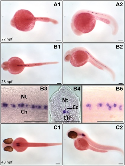
urp1 mRNA is restricted to the ventral spinal cord and hindbrain at early stages of development in zebrafish. Expression of urp1 revealed by ISH (BM purple, violet) on nacre embryos at 22 (A), 28 (B) and 48 hpf (C). At 22 hpf and 28 hpf, urp1+ cells occur only in the spinal cord at the level of the lateral floor plate (A, B), while from 48 hpf, they are mainly visible in the hindbrain (C). A1, B1, B5 and C1, dorsal views; A2, B2, B3 and C2, lateral views with dorsal up; B4, coronal section with dorsal up; all embryos oriented anterior left; B3 and B5 are details at higher magnifications of B2 and B1, respectively. Ch, chord; Cc, central canal; Nt, neural tube; Scale bar: 100 µm. EXPRESSION / LABELING:
|
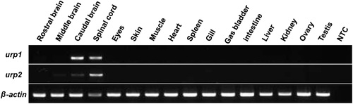
urp1 and urp2 mRNAs are exclusively detected in the brain and spinal cord in adult zebrafish. Tissue distribution of urp1 and urp2 mRNAs assessed by RT-PCR. Parallel amplification of zebrafish β-actin mRNA served as internal control. NTC, non-template control. EXPRESSION / LABELING:
|
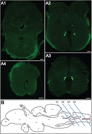
urp1 mRNA is found in the caudal part of the hindbrain in adult zebrafish. Expression of urp1 revealed by fluorescent ISH (FITC, green) on coronal sections of adult brain (A). urp1 mRNA is visible in neurons located in the intermediate reticular formation (A1) and the region of the glossopharyngeal-vagal motor nerve nuclei (A2–A3). More caudally, at the level of the junction between hindbrain and spinal cord, urp1 mRNA occurs at the ventral edge of the central canal (A4). Schematic sagittal view of an adult zebrafish brain depicting the distribution of urp1 mRNA (red dots). Levels of sections shown in A are indicated. The anatomical structures are designated according to [38] (B). CC, cerebellar crest; C, central canal; CCe, corpus cerebelli; DON, dorsal octavolateralis nucleus; EW, Edinger-Westphal nucleus; FLo, facial lobe; Ha, habenula; H, hypothalamus; IMRF, intermediate reticular formation; MO, medulla oblongata; NC, commissural nucleus of Cajal; nIX-X, glossopharyngeal-vagal motor nerve nuclei; OB, olfactive bulbs; OC, optic chiasma; P, pallium; PN, preopic nucleus; RV, rhombencephalic ventricle; SCsm, spinal cord somatomotor neurons; SP, subpallium; T, thalamus; TO, tectum opticum; TL, torus longitudinalis; TP, posterior tuberculum; TS, torus semicircularis; VLo, vagal lobe. Scale bars: 100 µm. EXPRESSION / LABELING:
|
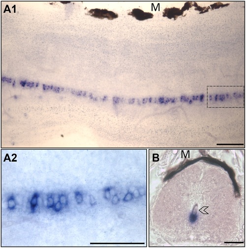
urp1 mRNA occurs in cells located along the ventral edge of the central canal of spinal cord in adult zebrafish. Expression of urp1 revealed by ISH (BM purple, violet) on free-floating sections of adult spinal cord. urp1+ cells form a quasi-continuous line at the ventral edge of the central canal (A). urp1+ cells are in close contact to the lumen of the central canal (arrowhead) (B). A1 and A2, lateral sections with dorsal up; B, coronal section with dorsal up. urp1+ cells boxed in A1 are shown in A2 at higher magnification. M, melanocytes. Scale bars: 50 µm. EXPRESSION / LABELING:
|
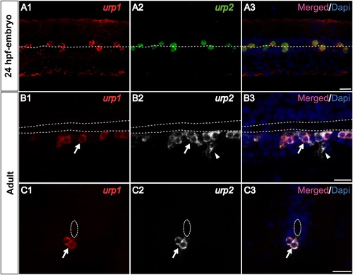
urp1 and urp2 mRNAs are mainly coexpressed in the same spinal cord cells in zebrafish. Simultaneous expression of urp1 and urp2 revealed by double fluorescent ISH (TAMRA, red for urp1, FITC, green for urp2 and DAPI in blue) on 24 hpf-embryo (A) and adult spinal cord sections (B, C). In embryo, all the stained cells contain both urp1 and urp2 mRNAs (A). In adult, although most of the stained cells are doubly-positive for urp1 and urp2 (arrow), some of the urp2+ cells are devoid of any urp1 mRNA (arrowhead). The white dash line indicates the central canal. A, dorsal view; B, sagittal section with dorsal up; C, coronal section with dorsal up. Scale bars: 15 µm. |
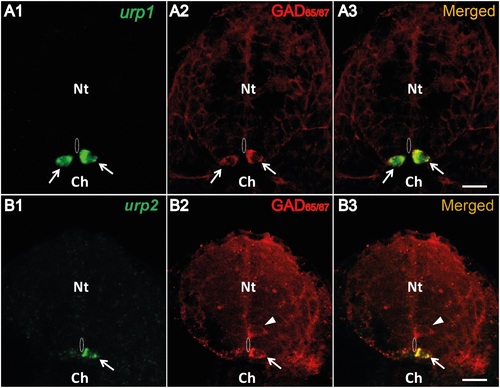
Both urp1+ and urp2+ cells are GABAergic neurons in the zebrafish embryo. urp1 (A) and urp2 (B) expression revealed by fluorescent ISH (FITC, green) on 24 hpf-embryo, together with a fluorescent immunostaining for GAD65/67 (Alexa Fluor 546, red). Both urp1+ and urp2+ cells are GAD+ (arrows). Note that only ventral KA (KA′′) cells are doubly stained. In contrast, dorsal KA (KA′) cells are GAD+ but do not express urp1 (arrowhead). The white dash line indicates the central canal. A and B, coronal sections with dorsal up. Scale bars: 15 µm. EXPRESSION / LABELING:
|
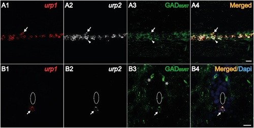
Both urp1+ and urp2+ cells in the spinal cord are GABAergic neurons in adult zebrafish. Simultaneous expression of urp1 and urp2 revealed by double fluorescent ISH (TAMRA, red for urp1 and Cy5, white for urp2) in adult spinal cord, coupled to a fluorescent immunostaining for GAD65/67 (Alexa Fluor 488, green) (A, B). Both urp1+ and urp2+ cells are GAD+ (A, B). Arrows designate triple-stained cells (A, B). Note the occurrence of some doubly-positive cells (urp2/GAD) that do not contain any urp1 (arrowhead) (A). Asterisks designate GABAergic interneurons located at the dorsal part of the spinal cord. The white dash line indicates the central canal. A, sagittal section with dorsal up; B, coronal section with dorsal up. Scale bars: 15 µm. EXPRESSION / LABELING:
|
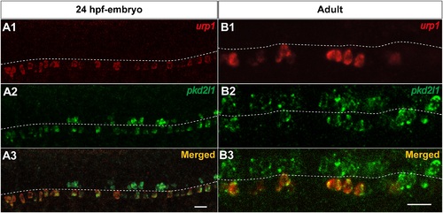
urp1+ cells express pkd2l1, a specific marker of spinal cerebrospinal fluid- contacting neurons in zebrafish. Simultaneous expression of urp1 and pkd2l1 revealed by double fluorescent ISH (TAMRA, red for urp1 and FITC, green for pkd2l1) on 24 hpf-embryo (A) and adult spinal cord sections (B). pkd2l1 mRNA is distributed in two rows of cells along the rostro-caudal axis of the spinal cord both in embryo and adult (A2, B2). All the urp1+ cells are pkd2l1+ (A1,3, B1,3) but only the ventral pkd2l1+ cells are urp1+. The white dash line indicates the central canal. A, lateral views; B, sagittal sections with dorsal up. Scale bars: 20µm. |
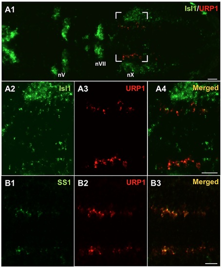
urp1+ cells express isl1 and ss1, two markers of the vagus motor nucleus in zebrafish embryo. Simultaneous expression of urp1 and isl1 (A) or ss1 (B) revealed by double fluorescent ISH (TAMRA, red for urp1 and FITC, green for isl1 or ss1) on 48 hpf-embryo. urp1+ cells are located at the level of the medial motor nucleus of the vagus. Most of them appear to be both isl1+ and ss1+. All pictures are dorsal views with anterior left. The boxed region in A1 is shown at higher magnification in A2–A4 and the same region is shown in B. nV, trigeminal nerve motor nuclei; nVII, facial nerve motor nuclei; nX, vagal nerve motor nuclei. Scale bars: 20 µm. EXPRESSION / LABELING:
|
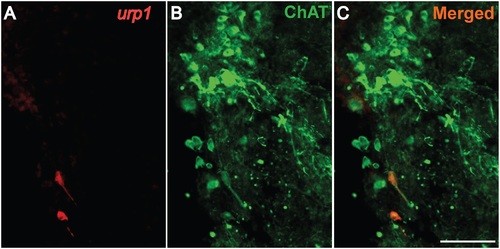
urp1+ cells in the hindbrain are cholinergic neurons expressing ChAT in adult zebrafish. urp1 expression revealed by fluorescent ISH (TAMRA, red) on coronal sections of adult brain, together with a fluorescent immunostaining for ChAT (Alexa 488, green). urp1+ cells express ChAT. Scale bars: 100 µm. EXPRESSION / LABELING:
|
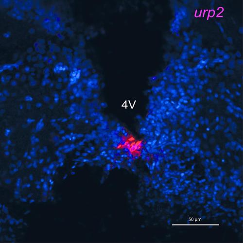
urp2 mRNA is found in cells contacting the fourth ventricle in the zebrafish adult brain. |
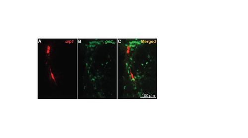
urp1+ cells in the hindbrain do not express gad67 in adult zebrafish. |