
FIGURE
Fig. 4
- ID
- ZDB-FIG-150504-29
- Publication
- Quan et al., 2015 - Comparative Distribution and In Vitro Activities of the Urotensin II-Related Peptides URP1 and URP2 in Zebrafish: Evidence for Their Colocalization in Spinal Cerebrospinal Fluid-Contacting Neurons
- Other Figures
- All Figure Page
- Back to All Figure Page
Fig. 4
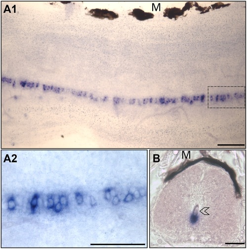
urp1 mRNA occurs in cells located along the ventral edge of the central canal of spinal cord in adult zebrafish. Expression of urp1 revealed by ISH (BM purple, violet) on free-floating sections of adult spinal cord. urp1+ cells form a quasi-continuous line at the ventral edge of the central canal (A). urp1+ cells are in close contact to the lumen of the central canal (arrowhead) (B). A1 and A2, lateral sections with dorsal up; B, coronal section with dorsal up. urp1+ cells boxed in A1 are shown in A2 at higher magnification. M, melanocytes. Scale bars: 50 µm. |
Expression Data
| Gene: | |
|---|---|
| Fish: | |
| Anatomical Term: | |
| Stage: | Adult |
Expression Detail
Antibody Labeling
Phenotype Data
Phenotype Detail
Acknowledgments
This image is the copyrighted work of the attributed author or publisher, and
ZFIN has permission only to display this image to its users.
Additional permissions should be obtained from the applicable author or publisher of the image.
Full text @ PLoS One