
Fig. 9
- ID
- ZDB-FIG-150504-34
- Publication
- Quan et al., 2015 - Comparative Distribution and In Vitro Activities of the Urotensin II-Related Peptides URP1 and URP2 in Zebrafish: Evidence for Their Colocalization in Spinal Cerebrospinal Fluid-Contacting Neurons
- Other Figures
- All Figure Page
- Back to All Figure Page
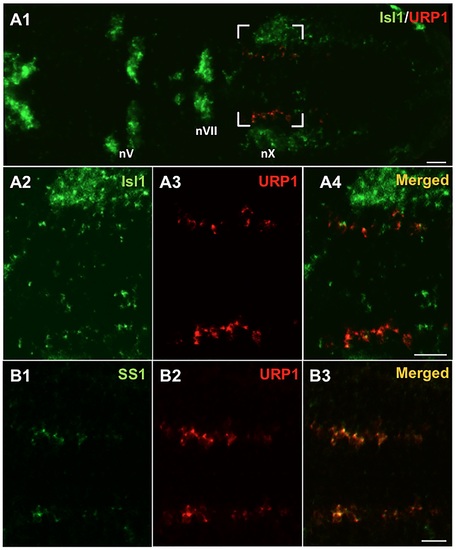
urp1+ cells express isl1 and ss1, two markers of the vagus motor nucleus in zebrafish embryo. Simultaneous expression of urp1 and isl1 (A) or ss1 (B) revealed by double fluorescent ISH (TAMRA, red for urp1 and FITC, green for isl1 or ss1) on 48 hpf-embryo. urp1+ cells are located at the level of the medial motor nucleus of the vagus. Most of them appear to be both isl1+ and ss1+. All pictures are dorsal views with anterior left. The boxed region in A1 is shown at higher magnification in A2–A4 and the same region is shown in B. nV, trigeminal nerve motor nuclei; nVII, facial nerve motor nuclei; nX, vagal nerve motor nuclei. Scale bars: 20 µm. |
| Genes: | |
|---|---|
| Fish: | |
| Anatomical Term: | |
| Stage: | Long-pec |