
Fig. 5
- ID
- ZDB-FIG-150504-30
- Publication
- Quan et al., 2015 - Comparative Distribution and In Vitro Activities of the Urotensin II-Related Peptides URP1 and URP2 in Zebrafish: Evidence for Their Colocalization in Spinal Cerebrospinal Fluid-Contacting Neurons
- Other Figures
- All Figure Page
- Back to All Figure Page
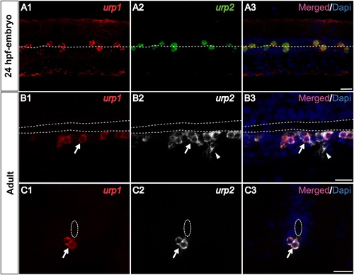
urp1 and urp2 mRNAs are mainly coexpressed in the same spinal cord cells in zebrafish. Simultaneous expression of urp1 and urp2 revealed by double fluorescent ISH (TAMRA, red for urp1, FITC, green for urp2 and DAPI in blue) on 24 hpf-embryo (A) and adult spinal cord sections (B, C). In embryo, all the stained cells contain both urp1 and urp2 mRNAs (A). In adult, although most of the stained cells are doubly-positive for urp1 and urp2 (arrow), some of the urp2+ cells are devoid of any urp1 mRNA (arrowhead). The white dash line indicates the central canal. A, dorsal view; B, sagittal section with dorsal up; C, coronal section with dorsal up. Scale bars: 15 µm. |
| Genes: | |
|---|---|
| Fish: | |
| Anatomical Term: | |
| Stage Range: | Prim-5 to Adult |