
Image
|
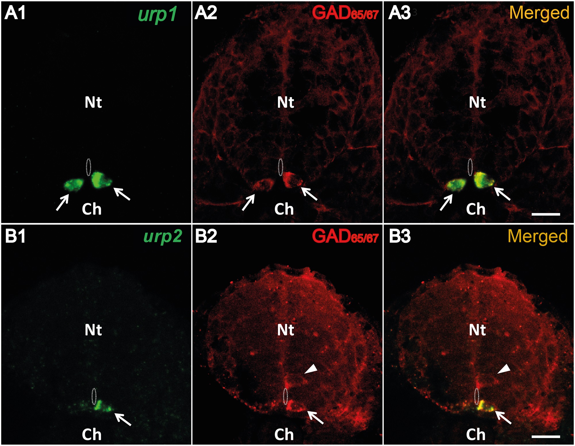
Figure Caption
Fig. 6 Both urp1+ and urp2+ cells are GABAergic neurons in the zebrafish embryo.
urp1 (A) and urp2 (B) expression revealed by fluorescent ISH (FITC, green) on 24 hpf-embryo, together with a fluorescent immunostaining for GAD65/67 (Alexa Fluor 546, red). Both urp1+ and urp2+ cells are GAD+ (arrows). Note that only ventral KA (KA′′) cells are doubly stained. In contrast, dorsal KA (KA′) cells are GAD+ but do not express urp1 (arrowhead). The white dash line indicates the central canal. A and B, coronal sections with dorsal up. Scale bars: 15 µm.
Figure Data
Acknowledgments
This image is the copyrighted work of the attributed author or publisher, and
ZFIN has permission only to display this image to its users.
Additional permissions should be obtained from the applicable author or publisher of the image.
Full text @ PLoS One