
|
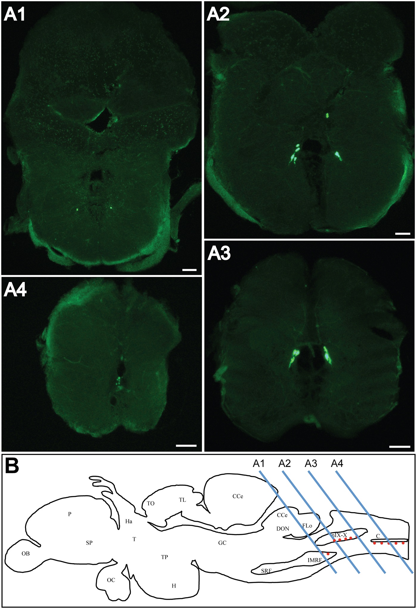
Fig. 3 urp1 mRNA is found in the caudal part of the hindbrain in adult zebrafish.
Expression of urp1 revealed by fluorescent ISH (FITC, green) on coronal sections of adult brain (A). urp1 mRNA is visible in neurons located in the intermediate reticular formation (A1) and the region of the glossopharyngeal-vagal motor nerve nuclei (A2–A3). More caudally, at the level of the junction between hindbrain and spinal cord, urp1 mRNA occurs at the ventral edge of the central canal (A4). Schematic sagittal view of an adult zebrafish brain depicting the distribution of urp1 mRNA (red dots). Levels of sections shown in A are indicated. The anatomical structures are designated according to [38] (B). CC, cerebellar crest; C, central canal; CCe, corpus cerebelli; DON, dorsal octavolateralis nucleus; EW, Edinger-Westphal nucleus; FLo, facial lobe; Ha, habenula; H, hypothalamus; IMRF, intermediate reticular formation; MO, medulla oblongata; NC, commissural nucleus of Cajal; nIX-X, glossopharyngeal-vagal motor nerve nuclei; OB, olfactive bulbs; OC, optic chiasma; P, pallium; PN, preopic nucleus; RV, rhombencephalic ventricle; SCsm, spinal cord somatomotor neurons; SP, subpallium; T, thalamus; TO, tectum opticum; TL, torus longitudinalis; TP, posterior tuberculum; TS, torus semicircularis; VLo, vagal lobe. Scale bars: 100 µm.