
|
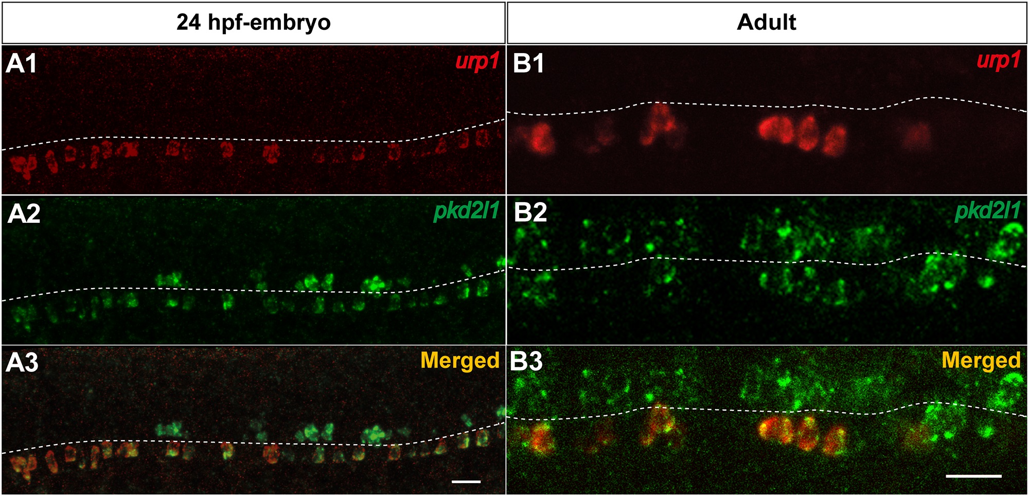
Fig. 8 urp1+ cells express pkd2l1, a specific marker of spinal cerebrospinal fluid- contacting neurons in zebrafish.
Simultaneous expression of urp1 and pkd2l1 revealed by double fluorescent ISH (TAMRA, red for urp1 and FITC, green for pkd2l1) on 24 hpf-embryo (A) and adult spinal cord sections (B). pkd2l1 mRNA is distributed in two rows of cells along the rostro-caudal axis of the spinal cord both in embryo and adult (A2, B2). All the urp1+ cells are pkd2l1+ (A1,3, B1,3) but only the ventral pkd2l1+ cells are urp1+. The white dash line indicates the central canal. A, lateral views; B, sagittal sections with dorsal up. Scale bars: 20µm.