
- Title
-
Dpysl2 (CRMP2) and Dpysl3 (CRMP4) phosphorylation by Cdk5 and DYRK2 is required for proper positioning of Rohon-Beard neurons and neural crest cells during neurulation in zebrafish
- Authors
- Tanaka, H., Morimura, R., and Ohshima, T.
- Source
- Full text @ Dev. Biol.
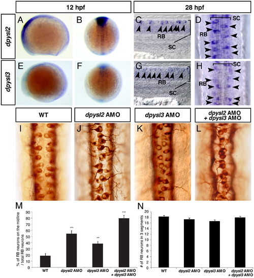
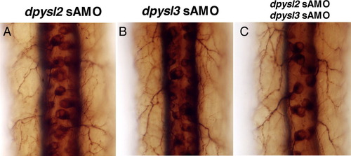
Loss of function of dpysl2 and dpysl3 inhibited the proper positioning of Rohon-Beard neurons in the dorsal part of the spinal cord. (A–H) Gene expression pattern of dpysl2 (A–D) and dpsyl3 (E–H) at 12 hpf (A, B, E, F) and at 28 hpf (C, D, G, H). Lateral view, anterior left (A, C, E, G) and dorsal view, anterior top (B, D, F, H). Arrowheads in C, D, G, H indicate RB neurons. (I–L) The position of RB neurons in the dorsal spinal cord in the wild-type (I) and the morphants (J–L) at 28 hpf. Dorsal view, anterior top. In the morphants, the position of RB neurons shifted medially compare to the wild-type embryos. (M, N) Quantitative data for the percentage of RB neurons on the midline (M), and total nomber of RB neurons within 3 segments (N). Data are mean ± SEM; NNP<0.01, NNNP<0.001, different from the wild-type embryos using 2-tailed paired Student′s t-test. EXPRESSION / LABELING:
PHENOTYPE:
|

The inhibition of phosphorylation of Dpysl2 and Dpysl3 caused the abnormal positioning of Rohon-Beard neurons. (A–F) The position of RB neurons in the dpysl2 morphants (A–C) and the dpysl3 morphants (D–F) which were co-injected with various AMO-resistant (5-mis) mRNAs at 28 hpf. Dorsal view, anterior top. The morphants which co-injected with mutant mRNAs showed severe positioning defects in RB neurons (B, C, E, F) compare to the normal mRNA co-injected morphants (A, D). (G) Quantitative data for the percentage of RB neurons on the midline. Data are mean ± SEM; NNP<0.01, NNNP<0.001, different from the normal mRNA co-injected morphants using 2-tailed paired Student′s t-test. |
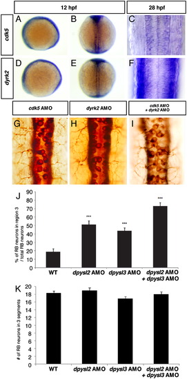
Loss of function of cdk5 and dyrk2 inhibited the proper positioning of Rohon-Beard neuron in the dorsal part of the spinal cord. (A–D) Gene expression pattern of cdk5 (A–C) and dyrk2 (D–F) at 12 hpf (A, B, D, E) and at 28 hpf (C, F). Lateral view, anterior left (A, D) and dorsal view, anterior top (B, C, E, F). (G–I) The position of RB neurons in the dorsal spinal cord of each morphant at 28 hpf. Dorsal view, anterior top. In the morphants, the position of RB neurons shifted medially compare to the wild-type embryos (see Fig. 1I). (J, K) Quantitative data for the percentage of RB neurons on the midline (J), and total nomber of RB neurons within 3 segments (K). Data are mean ± SEM; NNNP<0.001, different from the wild-type embryos using 2-tailed paired Student′s t-test. EXPRESSION / LABELING:
PHENOTYPE:
|
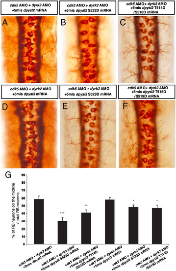
The abnormal position of Rohon-Beard neurons caused by loss of function of cdk5 and dyrk2 was rescued by phosphorylation-mimic forms of Dpysl2 and Dpysl3. (A–F) The position of RB neurons in the cdk5 and dyrk2 double morphants which co-injected with normal AMO-resistant (5-mis) dpysl2 or dpysl3 mRNAs (A, D) or with phosphorylation-mimic forms of dpysl2 or dpysl3 mRNAs (B, C, E, F) at 28 hpf. Dorsal view, anterior top. The morphants which co-injected with normal mRNAs showed severe positioning defects in RB neurons (A, D), however, phosphorylation-mimic forms of dpysl2 or dpysl3 mRNAs injected morphants partially recovered their phenotypes (B, C, E, F). (G) Quantitative data for the percentage of RB neurons on the midline. Data are mean ± SEM; NP<0.05, NNP<0.01, NNNP < 0.001, different from the normal mRNA co-injected morphants using 2-tailed paired Student′s t-test. |
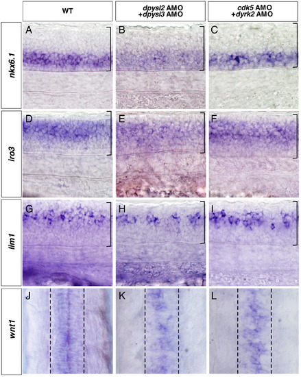
Loss of function of cdk5, dyrk2, dpysl2, and dpysl3 specifically affected the patterning of the dorsal-most part of the spinal cord. (A–L) Expression of nkx6.1 (A–C), iro3 (D–F), lim1 (G–I), and wnt1 (J–L) in the wild-type embryos (A, D, G, J), the dpysl2 and dpysl3 double morphants (B, E, H, K), and the cdk5 and dyrk2 double morphants (C, F, I, L) at 28 hpf, anterior left (A–I), anterior top (J–L). In the wild-type embryos, nkx6.1, iro3, and lim1 were expressed in the ventral, middle to dorsal, and dorsal part of the spinal cord, respectively (A, D, G). In the dorsal-most part of the spinal cord, wnt1 was expressed around the midline (J). In the dpysl2 and dpysl3 double morphants or the cdk5 and dyrk2 double morphants, the expression patterns of nkx6.1, iro3, and lim1 were similar to those of the wild-type embryos (B, C, E, F, H, I). However, the expression domain of wnt1 was abnormally distributed around the midline (K, L). Brackets in A–I and broken lines in J–L indicate the spinal cord. EXPRESSION / LABELING:
|
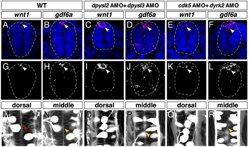
The dorsal midline structure was specifically affected by loss of function of cdk5, dyrk2, dpysl2 and dpysl3. (A–L) Expression of wnt1 and gdf6a in the wild-type embryos (A, B, G, H), the dpysl2 and dpysl3 double morphants (C, D, I, J), the cdk5 and dyrk2double morphants (E, F, K, L) at 28 hpf, transverse section, dorsal top, broken lines indicate margin of spinal cord. In the wild-type embryos, expression of wnt1 and gdf6a were restricted in the dorsal midline (A, B, G, H, arrow heads), while both genes expressed broader in the double morphants (C–F, I–L, arrow heads). (M–R) Dorsal view of spinal cord in the wild-type (M, N), the dpysl2 and dpysl3 double morphants (O, P), the cdk5 and dyrk2double morphants (Q, R) at 28 hpf. RB neurons and other interneurons were visualized using CREST2: GFP transgenic strain. Cell membrane was visualized using BODIPY-FL C5-ceramide. The dorsal midline border in the wild-type embryos (M, red arrowhead) was not observed in both double morphants (O, Q) while the apical surface in the middle part of spinal cord was normal like the wild-type embryos (N, P, R, yellow arrowheads). EXPRESSION / LABELING:
|
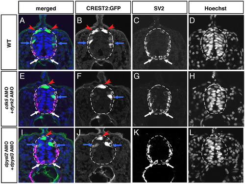
The position of Rohon-Beard neurons was specifically affected by loss of function of cdk5, dyrk2, dpysl2 and dpysl3. (A–L) The transverse views of the spinal cord in the wild-type embryos (A–D), the cdk5 and dyrk2 double morphants (E–H) and the dpysl2 and dpysl3 double morphants (I–L) at 28 hpf, dorsal top. Red arrowheads indicate RB neurons, blue arrows indicate interneurons and white allows indicate primary motor neurons. Broken lines indicate margin of spinal cord. In the wild-type embryos, CREST2:GFP-labeled RB neurons and interneurons (A, B), and SV2-labeled primary motor neurons (A, C) were located bilaterally. However, in the cdk5 and dyrk2 double morphants or the dpysl2 and dpysl3 double morphanst, RB neuron was observed on the midline while other types of neurons were located normally like the wild-type embryos (E–G, I–K). |
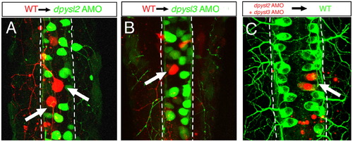
Dpysl2 and Dpysl3 non-cell autonomously affected the position of Rohon-Beard neurons. (A, B) The distribution of the transplanted wild-type RB neurons in the dpysl2 or dpysl3 morphants at 28 hpf, dorsal view, anterior top. In the dpysl2 morphant, the rhodamine dextran–labeled wild-type RB neurons were observed close to the midline (A, arrows). Similarly, in the dpysl3 morphant, the transplanted wild-type RB neurons were located in the relatively medial region, similar to those observed in the host morphant (B, arrow). (C) The distribution of the transplanted dpysl2 and dpysl3 morphant RB neurons in the wild-type embryos at 28 hpf, dorsal view, anterior top. The transplanted morphant RB neurons were located in the lateral part of the spinal cord, similar to those observed in the host embryos (C, arrow). Broken lines indicate the margin of the spinal cord. |
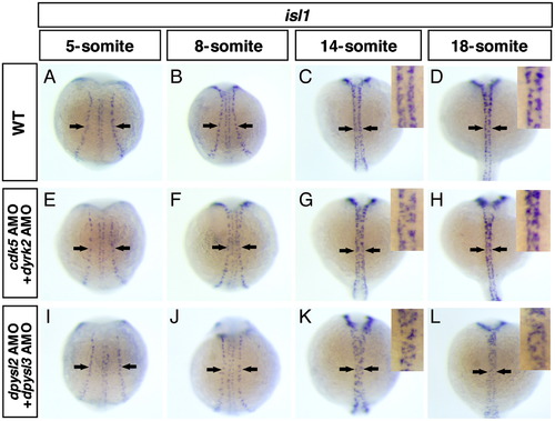
cdk5, dyrk2, dpysl2 and dpysl3 were involved in cell position during neurulation of the spinal cord but not in the formation of proneural domain. (A–L) The distribution of isl1-positive primary neurons in the wild-type embryos (A–D), the cdk5 and dyrk2 double morphants (E–H) and dpsyl2 and dpysl3 double morphants (I–L) through 5-somite to 18-somite stages, dorsal view, anterior top. Arrows indicate RB neurons, insets show high magnification views of the spinal cord. In the wild-type embryos, RB neurons were born in the lateral edge of neural plate and form the proneural domain (A). According to the neural keel and the neural rod formation, they migrated medially (B, C), and finally, they aligned bilaterally along the midline (D). In the double morphants, the proneural domains were normally formed (E, I). However, during the neurulation, RB neurons overshot their original position and some of them were reached to the midline (G, H, K, L). EXPRESSION / LABELING:
PHENOTYPE:
|
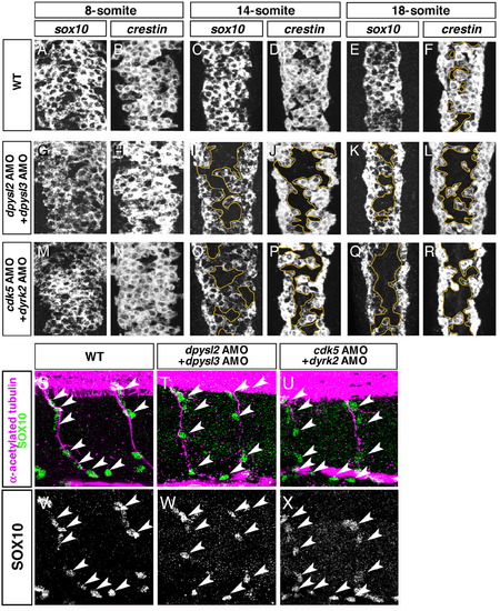
Cdk5, dyrk2, dpysl2, and dpysl3 were involved in the positioning of neural crest cells during neurulation of the spinal cord. (A–R) The expression of sox10 and crestin in the anterior part of the dorsal spinal cord in wild-type embryos (A–F), the dpysl2 and dpysl3 double morphants (G–L), and the cdk5 and dyrk2 double morphants (M–R) at 8- to 18-somite stages. In wild-type embryos, the sox10-positive or the crestin-positive NCCs distributed over the entire dorsal part of the spinal cord through the 8- to 18-somite stages (A–F). In the dpysl2 and dpysl3 double morphants, the sox10-positive or the crestin-positive NCCs also distributed throughout the dorsal part of the spinal cord at the 8-somite stage (G, H). However, at the 14- and 18-somite stages, the number of NCCs was reduced in the medial part of the spinal cord (I–L, surrounded by yellow line). Similarly, in the cdk5 and dyrk2 double morphants, a significant reduction in the number of NCCs was also observed in the medial part of the spinal cord (O–R, surrounded by yellow line). (S–X) Distribution of SOX-10-positive NCCs (arrow heads) along the axons of caudal primary motor neurons (CaP) in the wild-type embryos (S, V), the dpysl2 and dpysl3 double morphants (T, W), and the cdk5 and dyrk2 double morphants (U, X) at 36 hpf. EXPRESSION / LABELING:
PHENOTYPE:
|
|
Reprinted from Developmental Biology, 370(2), Tanaka, H., Morimura, R., and Ohshima, T., Dpysl2 (CRMP2) and Dpysl3 (CRMP4) phosphorylation by Cdk5 and DYRK2 is required for proper positioning of Rohon-Beard neurons and neural crest cells during neurulation in zebrafish, 223-236, Copyright (2012) with permission from Elsevier. Full text @ Dev. Biol.