
|
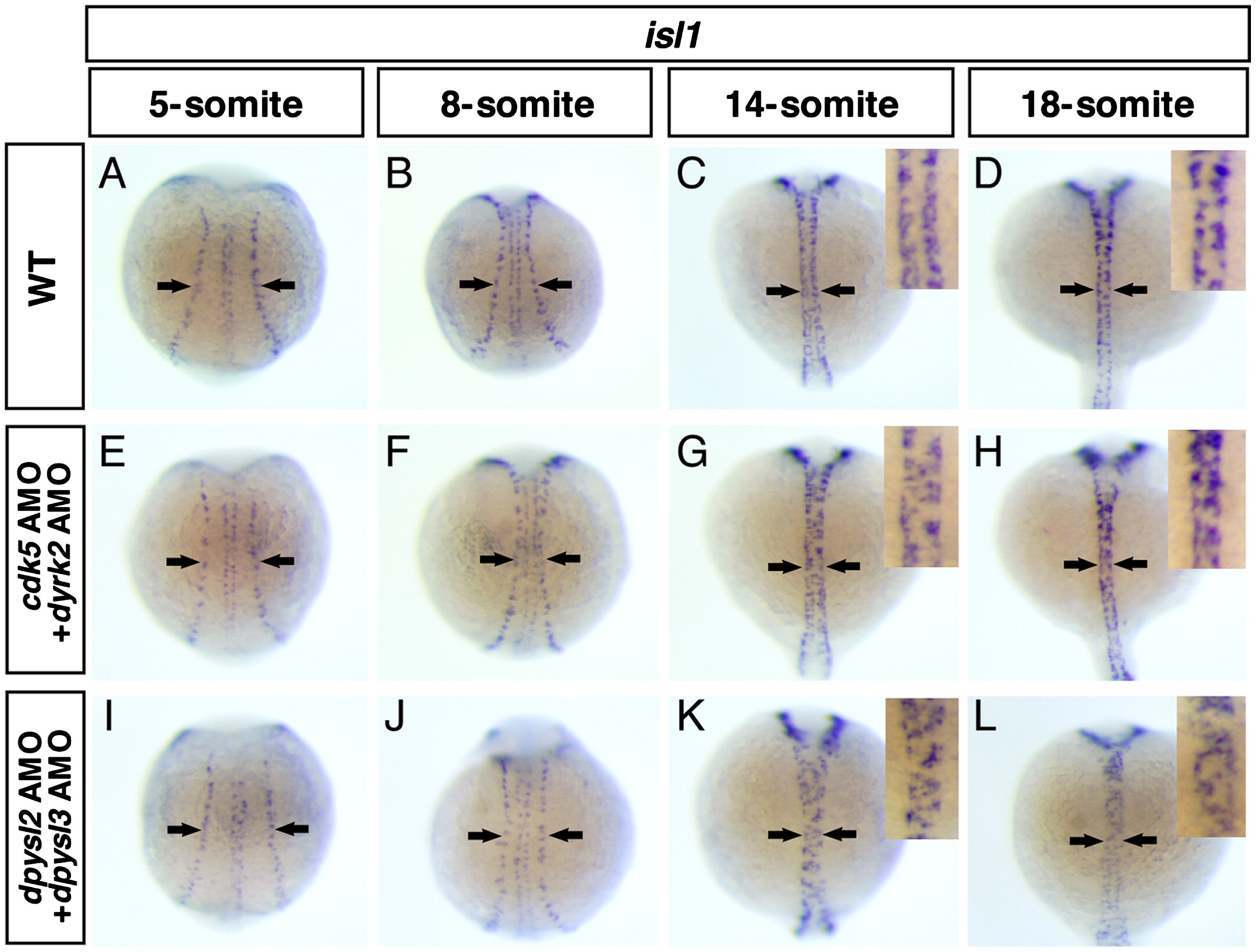
Fig. 9 cdk5, dyrk2, dpysl2 and dpysl3 were involved in cell position during neurulation of the spinal cord but not in the formation of proneural domain. (A–L) The distribution of isl1-positive primary neurons in the wild-type embryos (A–D), the cdk5 and dyrk2 double morphants (E–H) and dpsyl2 and dpysl3 double morphants (I–L) through 5-somite to 18-somite stages, dorsal view, anterior top. Arrows indicate RB neurons, insets show high magnification views of the spinal cord. In the wild-type embryos, RB neurons were born in the lateral edge of neural plate and form the proneural domain (A). According to the neural keel and the neural rod formation, they migrated medially (B, C), and finally, they aligned bilaterally along the midline (D). In the double morphants, the proneural domains were normally formed (E, I). However, during the neurulation, RB neurons overshot their original position and some of them were reached to the midline (G, H, K, L).
Reprinted from Developmental Biology, 370(2), Tanaka, H., Morimura, R., and Ohshima, T., Dpysl2 (CRMP2) and Dpysl3 (CRMP4) phosphorylation by Cdk5 and DYRK2 is required for proper positioning of Rohon-Beard neurons and neural crest cells during neurulation in zebrafish, 223-236, Copyright (2012) with permission from Elsevier. Full text @ Dev. Biol.