- Title
-
Chondrogenic and Gliogenic Subpopulations of Neural Crest Play Distinct Roles during the Assembly of Epibranchial Ganglia
- Authors
- Culbertson, M.D., Lewis, Z.R., and Nechiporuk, A.V.
- Source
- Full text @ PLoS One
|
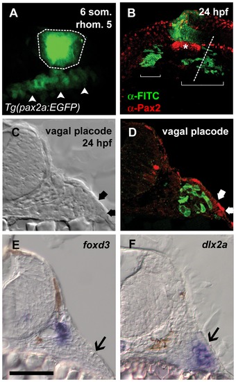
Neural crest cells migrate to abut epibranchial placodes. (A) Lateral view of Tg(pax2a:EGFP)w37/+ embryo at the six somite stage showing fluorescein uncaged in rhombomere 5 (outlined region). Placode precursor field expressing EGFP is visible below the uncaged region (arrowheads). (B) Lateral view of the same embryo at 24 hpf. Fluorescein-labeled NC cells derived from rhombomere 5 (green) are visible in two regions (brackets) ventral to the otic vesicle (asterisk). (C and D) Transverse section through embryo at level indicated by dotted line in B. Placode is visible as thickened ectoderm under transmitted light and by α-Pax2 staining in red (C and D, thick arrows). In D, fluorescein-labeled cells are just medial to, but excluded from, the placode. (E and F) In situ hybridization at the level of the vagal placode shows neural crest-derived glial precursors adjacent to the neural tube expressing foxd3; in F, a group of cells corresponding to the fluorescein-labeled region in D expressing the chondrogenic crest marker dlx2a. Placodes in E and F indicated by black arrows. Scale bar = 50 μm. EXPRESSION / LABELING:
|
|
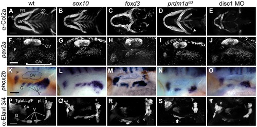
Disruptions in neural crest cause late defects in epibranchial ganglion morphology without affecting placode specification. (A–E) Ventral view of head skeleton visualized with anti-Collagen2a antibody at 4 dpf. (A) The following structures are visible in wildtype: Meckel′s cartilage, palatoquadrate, ceratohyal and ceratobranchial arches. Sox10 mutant (B) exhibits normal morphology. Radical disruption of structure is apparent in foxd3 mutants (C) and disc1 morphants (E). prdm1a mutant (D) displays grossly normal anterior jaw structures and deficits in the posterior arches; in this example, an unpaired ceratobranchial vestige can be seen (arrowhead). (F–J) Anti-Pax2 immunofluorescence at 24 hpf. Wildtype (F) shows facial placode and unsegmented glossopharyngeal/vagal placode field (K–O) phox2b expression at 48 hpf. Wildtype (K) displays normal segregation of facial,glossopharyngeal, and vagal ganglia. Defects are apparent in foxd3 (M) and prdm1a (N) mutants and disc1 morphant (O). sox10 mutant (K) display morphology and segmentation comparable to wildtype siblings (F). P–T: Ganglion morphology visualized by anti-Elavl3/4 antibody staining at 4 dpf. (K) Wildtype control shows glossopharyngeal and vagal ganglia. Posterior lateral line, statoacoustic and the fused trigeminal, anterior lateral line and facial ganglia can also be seen. Defects are apparent in sox10 (Q), foxd3 (R), prdm1a (S) mutants and disc1 morphant (T). Arrow in (S) shows an example of fusion often seen in prdm1a mutants. Abbreviations: mk = Meckel′s cartilage; pq = palatoquadrate; ch = ceratohyal; cb = ceratobranchial arches; OV = otic vesicle; F = facial ganglion; Tg/aLL/F = trigeminal/anterior lateral line/facial ganglion complex; SA = statoacoustic ganglion; pLLg = posterior lateral line ganglion; G = glossopharyngeal ganglion; V = vagal ganglia. Scale bar in (A) = 100 μm. Scale bars in (F), (K) and (P) = 50 μm. EXPRESSION / LABELING:
|
|
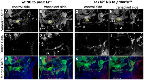
Wildtype and sox10 -/- NC is capable of rescuing cranial ganglion formation in prdm1anl3 mutant embryos. (A–F) EB ganglion rescue mediated by transplantation of wildtype NC into prdm1anl3 mutant embryos expressing Tg(NeuroD:EGFP). (A, B) Left (control) and right (transplanted) lateral views of prdm1anl3 mutant head with rhodamine-positive wildtype NC donor cells at 3 dpf. Tg(neuroD:EGFP)nl1 expression reveals an absence of identifiable cranial ganglion structures on the non-transplanted side (A) while transplanted side shows restoration of these structures, including identifiable glossopharyngeal and vagal ganglia. (C, D) Distribution of rhodamine-positive donor NC cells shows an absence of fluorescent label in the cranial ganglion field of the non-transplanted side (C). In the transplant side (D), rhodamine-positive donor cells are present in parallel columns extending ventrally from the ganglion field (arrows) and in a single line perpendicular and immediately dorsal to columns (star), likely corresponding to the ceratobranchials and the rostral basicranial commisure, respectively. (E and F) Merged channel high-magnification views of control (E) and rescue (F) sides with DAPI counterstain (blue). Note DAPI-labeled branchial arches containing rhodamine-positive donor cells (brackets). (G–L) Partial EB ganglion rescue following transplantation of sox10 -/- NC cells into prdm1anl3 host embryos. (G,H) Tg(NeuroD:EGFP) expression in prdm1anl3 embryo showing absence of small EB ganglia in control side (G) and a partial rescue of ganglion formation in ventral vagal region of transplanted side (arrowhead). (I,J) Distribution of rhodamine-labeled donor cells in control side (I) and rescue side (J). (K,L) Merged channel high-magnification views of control (K) and rescue (L) sides with DAPI counterstain. Rescued side shows juxtaposition of rescued ganglion with the rhodamine-positive arches (brackets). Margins of statoacoustic ganglia are indicated by dashed white lines. Abbreviations: Tg/aLL/F = trigeminal/anterior lateral line/facial ganglion complex; SA = statoacoustic ganglion; pLLg = posterior lateral line ganglion; G = glossopharyngeal ganglion; V = vagal ganglia. Scale bar in (A) = 50 μm. |
|
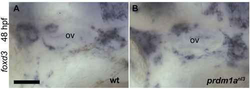
Glial NC cells are normal in the prdm1anl3 mutant. Lateral view showing that comparable foxd3 expression is detected by in situ hybridization in the region of the otic vesicle (ov) in wildtype (A) and prdm1anl3 mutant embryo (B) at 48 hpf. Abbreviation: OV = otic vesicle. Scale bar in (A) = 50 μm. EXPRESSION / LABELING:
|
|
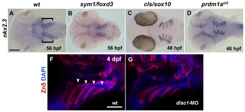
Endodermal pouches are present in NC-depleted mutants. (A–D) Ventral views showing nkx2.3 expression as detected by in situ hybridization in the head of wildtype (A) and foxd3 (B), sox10 (C) and prdm1a (D) mutants at 2 dpf. Endodermal pouches expressing nkx2.3 transcript are visible as bilateral sets of parallel linear expression domains posterior to the eyes (A, brackets). (E and F) Lateral view of Zn-5 immunofluorescence (red) marking endodermal pouches at 5 dpf in wildtype (E, arrowheads) and disc-1 morphant (F). Embryos were stained with DAPI (blue) to visualize nuclei. Scale bar in (A) = 100 μm. Scale bar in (E) = 50 μm. |
|
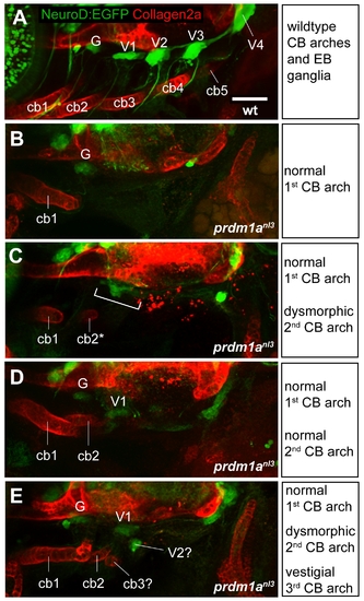
Relationship between CB arches and EB ganglia formation in prdm1anl3 mutants. (A) Composite lateral view of EB ganglia visualized with Tg(neuroD:EGFP)nl1 transgene (green) alongside cartilaginous CB arches stained for Collagen2a (red) in a 4 dpf wildtype larva. Fluoresence demonstrates spatial association of the first ceratobranchial arch with the glossopharyngeal ganglion and the second through fifth ceratobranchial arches with the first through fourth vagal ganglia, respectively. (B–E) Composite lateral views of prdm1anl3 mutant larvae exhibiting different degrees of CB arch formation. (B) A single, normally-formed first CB ventral to a cluster of EGFP-positive cells that corresponds to the glossopharyngeal ganglion. Note the absence of other small ganglia. (C) Normally-formed first CB arch alongside a dysmorphic second ceratobranchial arch. A fused ganglion structure is visible dorsal to the arches (bracket). (D) Normal formation of first and second CB arches. Distinct glossopharyneal and first vagal ganglia are positioned dorsal to the arch bodies. (E) Normally-formed first CB arch with dysmorphic second arch and presumptive third arch vestige. As in D, distinct glossopharygeal and first vagal ganglia are visible dorsal to arch structures. In addition, a small EGFP-positive cluster of cells can be seen between the arches and ganglia. Abbreviations: G = glossopharyngeal ganglion; V1–V4 = first through fourth vagal ganglia; cb1–cb5 = ceratobranchial arches 1–5. Scale bar = 50 μm. |
|
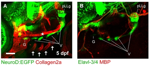
Spatial relationship between neurons, glia, and cartilage in the zebrafish head. (A) Lateral view of cranial ganglia in a 5 dpf wildtype larva expressing Tg(neurod:EGFP) and stained for Collagen2a (red). The rostral basicranial commisure (asterisk) can be seen positioned dorsally to the cranial ganglia. The Collagen2a-positive branchial arches are located ventrally (arrows). (B) Lateral view of cranial ganglia in a 5 dpf wildtype larva as visualized with immunofluoresence against Elavl-3/4 (green). Glial cells are stained with an antibody recognizing MBP (red). Scale bar in (A) = 50 μm. EXPRESSION / LABELING:
|

