
|
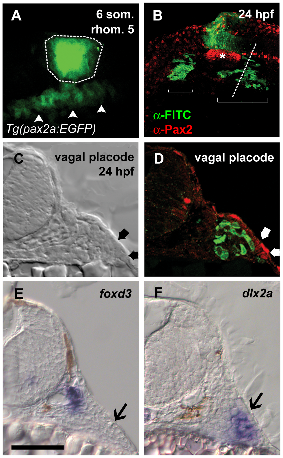
Fig. 1
Neural crest cells migrate to abut epibranchial placodes.
(A) Lateral view of Tg(pax2a:EGFP)w37/+ embryo at the six somite stage showing fluorescein uncaged in rhombomere 5 (outlined region). Placode precursor field expressing EGFP is visible below the uncaged region (arrowheads). (B) Lateral view of the same embryo at 24 hpf. Fluorescein-labeled NC cells derived from rhombomere 5 (green) are visible in two regions (brackets) ventral to the otic vesicle (asterisk). (C and D) Transverse section through embryo at level indicated by dotted line in B. Placode is visible as thickened ectoderm under transmitted light and by α-Pax2 staining in red (C and D, thick arrows). In D, fluorescein-labeled cells are just medial to, but excluded from, the placode. (E and F) In situ hybridization at the level of the vagal placode shows neural crest-derived glial precursors adjacent to the neural tube expressing foxd3; in F, a group of cells corresponding to the fluorescein-labeled region in D expressing the chondrogenic crest marker dlx2a. Placodes in E and F indicated by black arrows. Scale bar = 50 μm.