
Fig. 3
- ID
- ZDB-IMAGE-110928-20
- Publication
- Culbertson et al., 2011 - Chondrogenic and Gliogenic Subpopulations of Neural Crest Play Distinct Roles during the Assembly of Epibranchial Ganglia
- All Figures
- Figures for Culbertson et al., 2011
|
Fig. 3
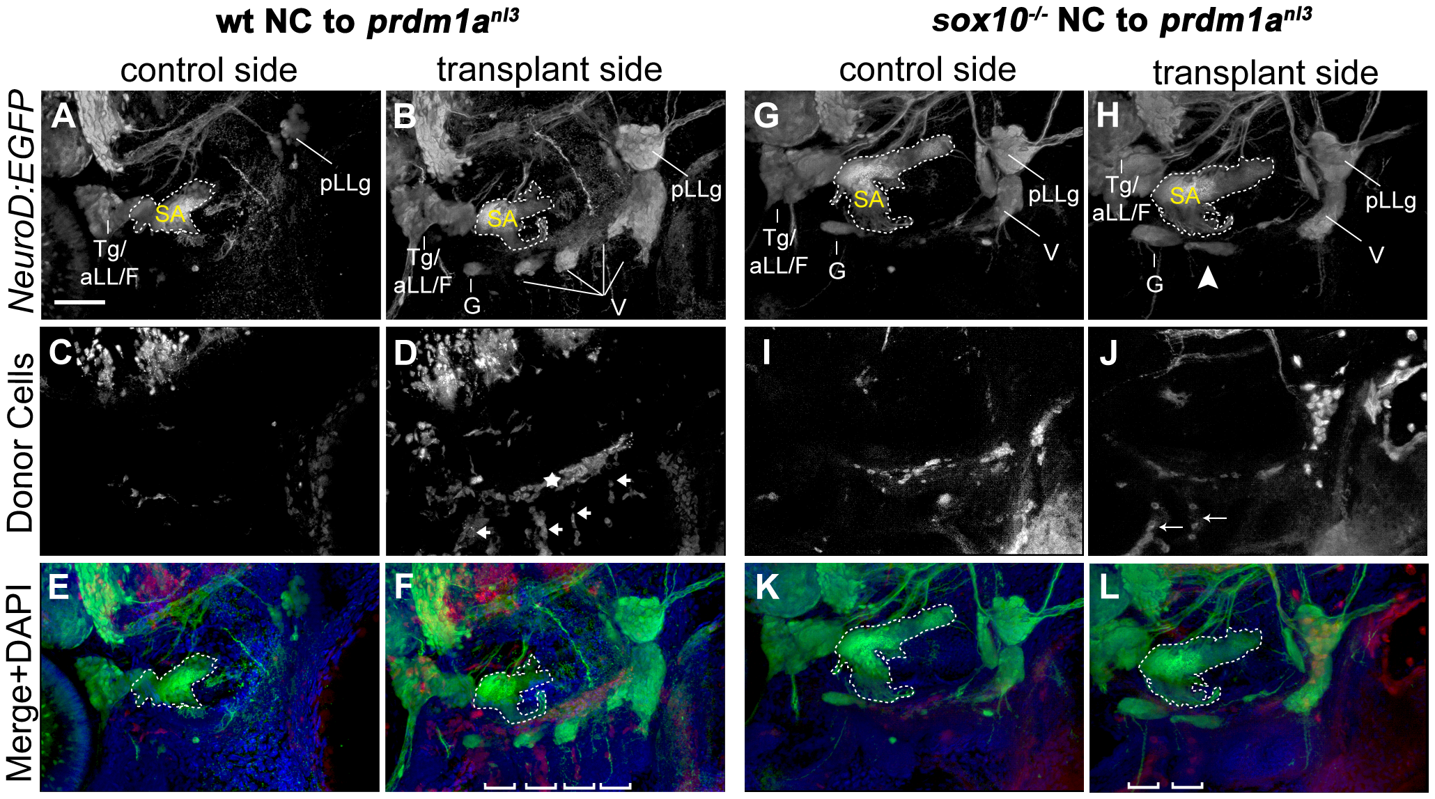
Wildtype and sox10 -/- NC is capable of rescuing cranial ganglion formation in prdm1anl3 mutant embryos.
(A–F) EB ganglion rescue mediated by transplantation of wildtype NC into prdm1anl3 mutant embryos expressing Tg(NeuroD:EGFP). (A, B) Left (control) and right (transplanted) lateral views of prdm1anl3 mutant head with rhodamine-positive wildtype NC donor cells at 3 dpf. Tg(neuroD:EGFP)nl1 expression reveals an absence of identifiable cranial ganglion structures on the non-transplanted side (A) while transplanted side shows restoration of these structures, including identifiable glossopharyngeal and vagal ganglia. (C, D) Distribution of rhodamine-positive donor NC cells shows an absence of fluorescent label in the cranial ganglion field of the non-transplanted side (C). In the transplant side (D), rhodamine-positive donor cells are present in parallel columns extending ventrally from the ganglion field (arrows) and in a single line perpendicular and immediately dorsal to columns (star), likely corresponding to the ceratobranchials and the rostral basicranial commisure, respectively. (E and F) Merged channel high-magnification views of control (E) and rescue (F) sides with DAPI counterstain (blue). Note DAPI-labeled branchial arches containing rhodamine-positive donor cells (brackets). (G–L) Partial EB ganglion rescue following transplantation of sox10 -/- NC cells into prdm1anl3 host embryos. (G,H) Tg(NeuroD:EGFP) expression in prdm1anl3 embryo showing absence of small EB ganglia in control side (G) and a partial rescue of ganglion formation in ventral vagal region of transplanted side (arrowhead). (I,J) Distribution of rhodamine-labeled donor cells in control side (I) and rescue side (J). (K,L) Merged channel high-magnification views of control (K) and rescue (L) sides with DAPI counterstain. Rescued side shows juxtaposition of rescued ganglion with the rhodamine-positive arches (brackets). Margins of statoacoustic ganglia are indicated by dashed white lines. Abbreviations: Tg/aLL/F = trigeminal/anterior lateral line/facial ganglion complex; SA = statoacoustic ganglion; pLLg = posterior lateral line ganglion; G = glossopharyngeal ganglion; V = vagal ganglia. Scale bar in (A) = 50 μm.