
Fig. S2
- ID
- ZDB-IMAGE-110928-22
- Genes
- Antibodies
- Publication
- Culbertson et al., 2011 - Chondrogenic and Gliogenic Subpopulations of Neural Crest Play Distinct Roles during the Assembly of Epibranchial Ganglia
- All Figures
- Figures for Culbertson et al., 2011
|
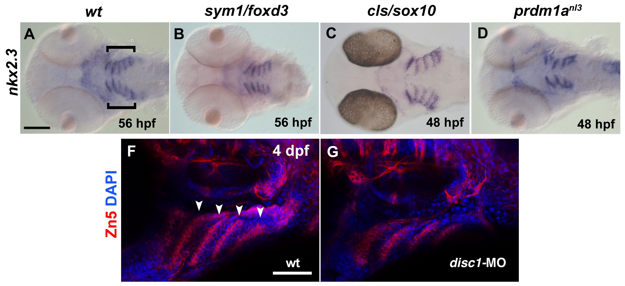
Fig. S2
Endodermal pouches are present in NC-depleted mutants. (A–D) Ventral views showing nkx2.3 expression as detected by in situ hybridization in the head of wildtype (A) and foxd3 (B), sox10 (C) and prdm1a (D) mutants at 2 dpf. Endodermal pouches expressing nkx2.3 transcript are visible as bilateral sets of parallel linear expression domains posterior to the eyes (A, brackets). (E and F) Lateral view of Zn-5 immunofluorescence (red) marking endodermal pouches at 5 dpf in wildtype (E, arrowheads) and disc-1 morphant (F). Embryos were stained with DAPI (blue) to visualize nuclei. Scale bar in (A) = 100 μm. Scale bar in (E) = 50 μm.