
- Title
-
Patterning of angiogenesis in the zebrafish embryo
- Authors
- Childs, S., Chen, J.-N., Garrity, D.M., and Fishman, M.C.
- Source
- Full text @ Development
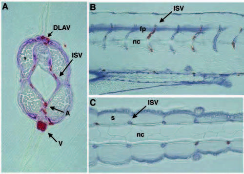
ISV relationships to somite, notochord and neural tube. (A) Cross section of posterior trunk shows close apposition of a pair of ISVs with somite, notochord and neural tube. (B) Sagittal section shows extremely regular pattern of ISVs closely associated with the somite boundary in the ventral trunk. Anterior is towards the left, and dorsal is upwards. (C) In this transverse section, pairs of ISVs are located at the somite boundaries that surround the notochord. Anterior is towards the left. Vessels are labeled by reaction of endogenous alkaline phosphatase. Blood is labeled with Isolectin B4. DLAV, dorsal longitudinal anastomotic vessel; ISV, intersegmental vessel; A, dorsal aorta; V, posterior cardinal vein; fp, floor plate; nc, notochord; s, somite. |
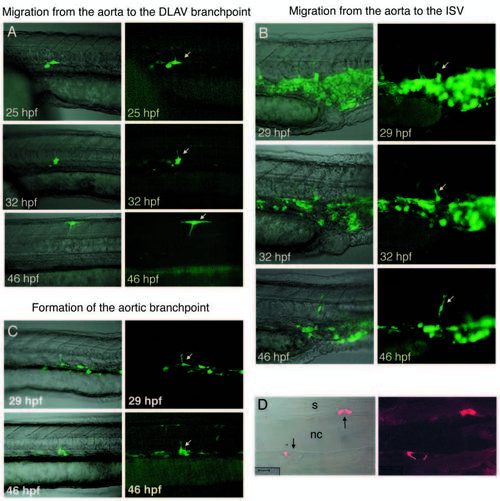
Time-lapse of angioblasts migrating from the aorta to assume the three stereotypical cell fates in the ISV: single cells expressing GFP under the tie2 promotor were photographed over a 21 hour period. Examples are shown of cells assuming the three positions. (A) T-shaped cell based in the DLAV projecting into the ISV. Arrows show the cell turning its migration; (B) ISV connector cell (arrow); and (C) aorta and ISV T-shaped cell (arrow). (D) Transverse sections of these tie2 GFP labeled embryos show that one to two cells (arrows) surround each ISV. In A-C, the right panel shows the fluorescent image and the left panel shows the fluorescent image superimposed on the phase contrast one. Anterior is towards the left, and dorsal is upwards. Scale bar: 10 μm. |
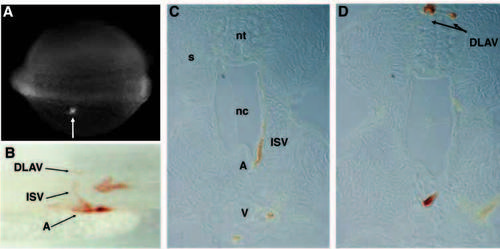
Lineage tracing of trunk shows ISVs derive from the LPM, via the axial vessels. (A) Fluorescein dye is uncaged by the laser in cells in the LPM of a nine somite embryo adjacent to somite nine. (B) Laser activation of cells in the LPM leads to labeling of aortic, connector and T-cells. (C) Cross section of a labeled ISV. (D) Cross section of ipsilateral and contralateral labeled DLAV cells. In A,B, anterior is towards the left. Dorsal is upwards. DLAV, dorsal longitudinal anastomotic vessel; ISV, intersegmental vessel; A, dorsal aorta; V, posterior cardinal vein; nt, neural tube; nc, notochord; s, somite. |
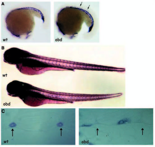
out of bounds mutant angioblasts migrate precociously, sprout from anomalous positions and generate abnormal patterns. (A) In situ hybridization of fli1 on 18 somite obd embryos demonstrates ectopic angioblast migration into the somite region in mutant embryos compared with wild type. Arrows denote ectopic cells. (B) The dorsal aorta and posterior cardinal vein are normally patterned as revealed by alkaline phosphatase staining in obd, while angiogenic vessels of the SIVs and ISVs are completely disorganized in the trunk region. (C) Transverse sections of alkaline phosphatase stained embryos demonstrate that ISVs are positioned at the intersegmental boundaries (arrows) in wild type, and are positioned randomly within the somites of obd mutants. EXPRESSION / LABELING:
PHENOTYPE:
|
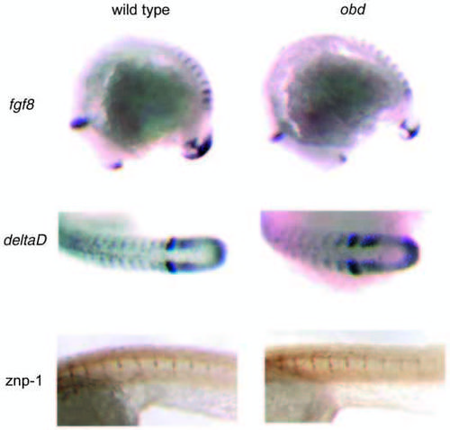
Anteroposterior patterning of the somite and primary motoneuron patterning are unaffected in obd mutants. In situ hybridization of wild-type zebrafish with probes for fgf8 or deltaD demonstrates a segmental expression pattern in the anterior somite. The expression pattern in obd homozygous mutant animals is identical to that in wild type. Antibody staining with znp-1 marks primary motoneurons in zebrafish. The pattern is identical in obd and wild-type embryos. EXPRESSION / LABELING:
|
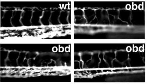
obd ISVs are patent but tortuous. Angiograms of the trunk region of wild-type and three obd mutants demonstrate the abnormal and variable pattern of patent obd mutant ISVs. The spacing of ISVs in obd appears most affected ventrally where the vessels originate, as opposed to dorsally where they terminate. PHENOTYPE:
|
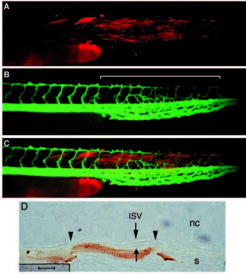
obd cells in ventral somite perturb wild-type endothelium: obd acts cell non-autonomously. (A) Transplanted obd somite labeled with rhodamine-dextran in the tail of an unlabeled wild-type embryo at 52 hpf. (B) Angiogram of the same embryo showing normally patterned ISVs in regions of no transplant (or where dorsal obd somite was transplanted) and vascular disorganization in the tail region where obd ventral somite was transplanted (bracket). (C) Overlay of A,B. (D) ISVs composed of wild-type endothelial cells grow mid-somite (borders of the somites are indicated by arrowheads), in the vicinity of rhodamine dextran biotin-labeled obd somite. ISV, intersegmental vessel; nc, notochord; s, somite. Scale bar is 10 μm. |
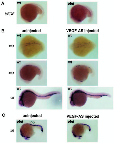
VEGF antisense delays precocious migration of obd angioblasts. (A) VEGF is normally expressed segmentally, beginning at the 17 somite stage, in both wild-type and obd embryos. (B) In a wild-type untreated embryo at 14 somites, angioblasts have migrated from the LPM to the midline while in a 1 mM VEGF-antisense treated embryo, tie1-positive cells are still localized laterally (top row). The dorsal aorta and vein are formed in both treated and untreated embryos at 19 somites, although the vessels in the injected embryo are discontinuous (middle row). At 24 hours, angiogenic sprouts are evident in an untreated embryo but are not seen in the treated embryos (bottom row). (C) The ectopic sprouts evident in a 19 somite obd embryo (arrows), are completely inhibited in the VEGF-AStreated obd mutant embryo. EXPRESSION / LABELING:
PHENOTYPE:
|