
|
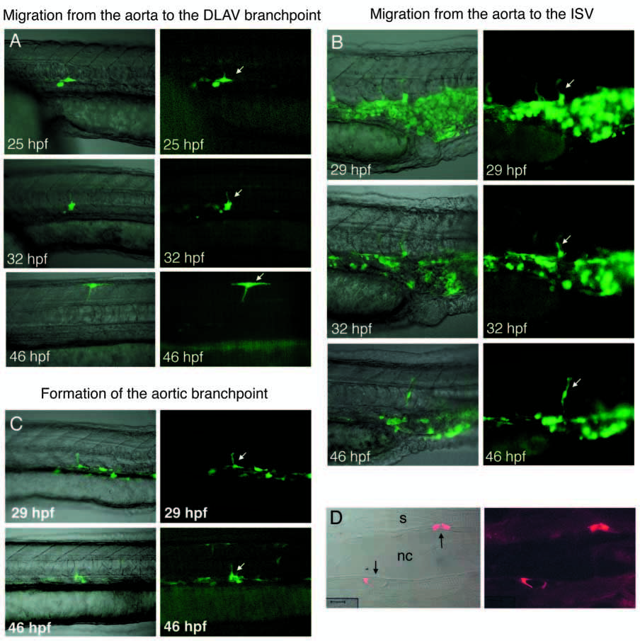
Fig. 2 Time-lapse of angioblasts migrating from the aorta to assume the three stereotypical cell fates in the ISV: single cells expressing GFP under the tie2 promotor were photographed over a 21 hour period. Examples are shown of cells assuming the three positions. (A) T-shaped cell based in the DLAV projecting into the ISV. Arrows show the cell turning its migration; (B) ISV connector cell (arrow); and (C) aorta and ISV T-shaped cell (arrow). (D) Transverse sections of these tie2 GFP labeled embryos show that one to two cells (arrows) surround each ISV. In A-C, the right panel shows the fluorescent image and the left panel shows the fluorescent image superimposed on the phase contrast one. Anterior is towards the left, and dorsal is upwards. Scale bar: 10 μm.