
Fig. 3
- ID
- ZDB-FIG-140326-18
- Publication
- Childs et al., 2002 - Patterning of angiogenesis in the zebrafish embryo
- Other Figures
- All Figure Page
- Back to All Figure Page
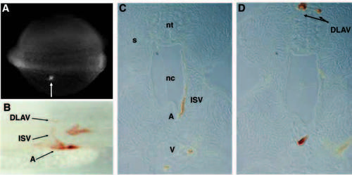
Lineage tracing of trunk shows ISVs derive from the LPM, via the axial vessels. (A) Fluorescein dye is uncaged by the laser in cells in the LPM of a nine somite embryo adjacent to somite nine. (B) Laser activation of cells in the LPM leads to labeling of aortic, connector and T-cells. (C) Cross section of a labeled ISV. (D) Cross section of ipsilateral and contralateral labeled DLAV cells. In A,B, anterior is towards the left. Dorsal is upwards. DLAV, dorsal longitudinal anastomotic vessel; ISV, intersegmental vessel; A, dorsal aorta; V, posterior cardinal vein; nt, neural tube; nc, notochord; s, somite. |