|
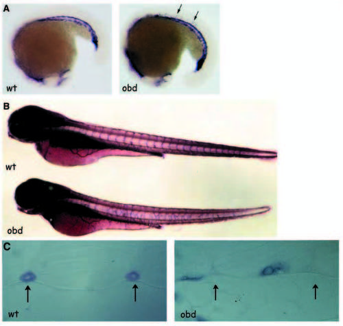
out of bounds mutant angioblasts migrate precociously, sprout from anomalous positions and generate abnormal patterns. (A) In situ hybridization of fli1 on 18 somite obd embryos demonstrates ectopic angioblast migration into the somite region in mutant embryos compared with wild type. Arrows denote ectopic cells. (B) The dorsal aorta and posterior cardinal vein are normally patterned as revealed by alkaline phosphatase staining in obd, while angiogenic vessels of the SIVs and ISVs are completely disorganized in the trunk region. (C) Transverse sections of alkaline phosphatase stained embryos demonstrate that ISVs are positioned at the intersegmental boundaries (arrows) in wild type, and are positioned randomly within the somites of obd mutants.
|