
Fig. 1
- ID
- ZDB-IMAGE-140326-9
- Publication
- Childs et al., 2002 - Patterning of angiogenesis in the zebrafish embryo
- All Figures
- Figures for Childs et al., 2002
|
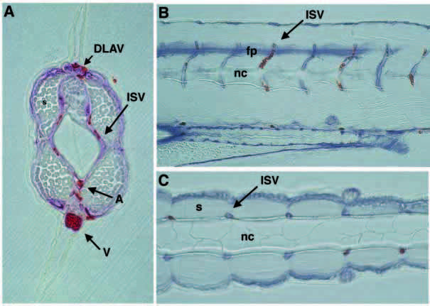
Fig. 1 ISV relationships to somite, notochord and neural tube. (A) Cross section of posterior trunk shows close apposition of a pair of ISVs with somite, notochord and neural tube. (B) Sagittal section shows extremely regular pattern of ISVs closely associated with the somite boundary in the ventral trunk. Anterior is towards the left, and dorsal is upwards. (C) In this transverse section, pairs of ISVs are located at the somite boundaries that surround the notochord. Anterior is towards the left. Vessels are labeled by reaction of endogenous alkaline phosphatase. Blood is labeled with Isolectin B4. DLAV, dorsal longitudinal anastomotic vessel; ISV, intersegmental vessel; A, dorsal aorta; V, posterior cardinal vein; fp, floor plate; nc, notochord; s, somite.