
Fig. 6
- ID
- ZDB-FIG-240731-76
- Publication
- Lee et al., 2024 - Mycb and Mych stimulate Müller glial cell reprogramming and proliferation in the uninjured and injured zebrafish retina
- Other Figures
- All Figure Page
- Back to All Figure Page
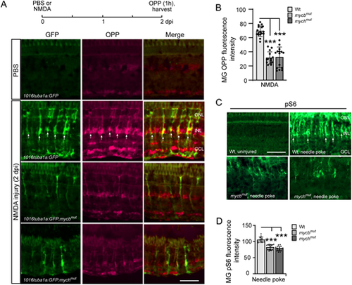
Injury-induced protein synthesis in reprogrammed MG is regulated by Mycb and Mych. (A) Experimental timeline is shown at the top. Representative images showing OPP-based protein synthesis detection in 1016 tuba1a:GFP, 1016 tuba1a:GFP;mycbmut and 1016 tuba1a:GFP;mychmut fish retinas at 2 days post-intravitreal injection of PBS or NMDA. Arrows point to GFP+/OPP+ co-stained cells, which are predominantly detected in 1016 tuba1a:GFP fish. (B) Quantification of data presented in A and additional experiments. (C) pS6 immunofluorescence on uninjured and injured (2 dpi) retinas from Wt, mycbmut and mychmut fish. Arrows point to representative pS6+ cells, which are predominantly detected in the injured fish retina. (D) Quantification of pS6 immunofluorescence in Wt, mycbmut and mychmut fish. The number of biological replicates (n) is indicated by the dots in each graph. ***P<0.001. dpi, days post injury; GCL, ganglion cell layer; INL, inner nuclear layer; ONL, outer nuclear layer; OPP, O-propargyl-puromycin. Scale bars: 100 μm. |