
Fig. 2
- ID
- ZDB-FIG-240731-72
- Publication
- Lee et al., 2024 - Mycb and Mych stimulate Müller glial cell reprogramming and proliferation in the uninjured and injured zebrafish retina
- Other Figures
- All Figure Page
- Back to All Figure Page
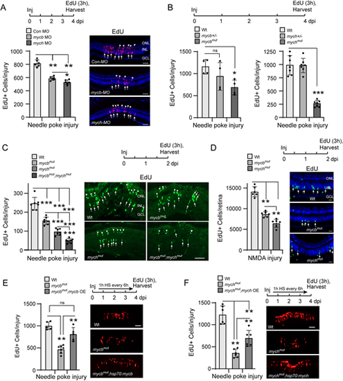
Mycb and Mych regulate MG proliferation in the injured retina. (A) Experimental timeline is shown at the top. Bar graph (left) and representative images (right) show the consequence of Mycb and Mych knockdown with antisense morpholino-modified oligonucleotide (MO) on MG proliferation (EdU+ cells, magenta, arrows) and rod progenitor proliferation (arrowheads in ONL) in needle poke-injured retina at 4 dpi. Retinal cells identified with DAPI (blue). (B) Experimental timeline is shown at the top. Bar graphs show the consequence of Mycb and Mych mutation on MG proliferation (EdU+ cells) in needle poke-injured retina at 4 dpi. (C) Experimental timeline is shown above the photomicrographs (right). Arrows point to proliferating MG in the INL and arrowheads point to proliferating rod progenitors in the ONL. Bar graph (left) and representative images (right) show the consequence of individual and combined Mycb and Mych mutation on MG proliferation (EdU+ cells; green) in the needle poke-injured retina at 2 dpi. (D) As in C, but NMDA was used to injure the retina. Retinal cells identified with DAPI (blue). (E,F) Experimental timeline is shown above the photomicrographs [arrow indicates 1 h heat shock (HS) repeated every 6 h]. Bar graph (left) and representative images (right) show that mycb OE rescues MG proliferation (EdU detection, red) in mycbmut;hsp70l:mycb fish (E), and mych OE only partially rescues MG proliferation in mychmut;hsp70l:mych fish (F). The number of biological replicates (n) is indicated by the dots in each graph. Error bars are s.d. *P<0.05, **P<0.01, ***P<0.001. dpi, days post-injury; GCL, ganglion cell layer; HS, heat shock; Inj, injury; INL, inner nuclear layer; ns, not significant; OE, overexpression; ONL, outer nuclear layer. Scale bars: 100 μm (A,C,E,F); 50 μm (D). |