
Figure 5
- ID
- ZDB-FIG-231205-5
- Publication
- Wilcockson et al., 2023 - An improved Erk biosensor detects oscillatory Erk dynamics driven by mitotic erasure during early development
- Other Figures
- All Figure Page
- Back to All Figure Page
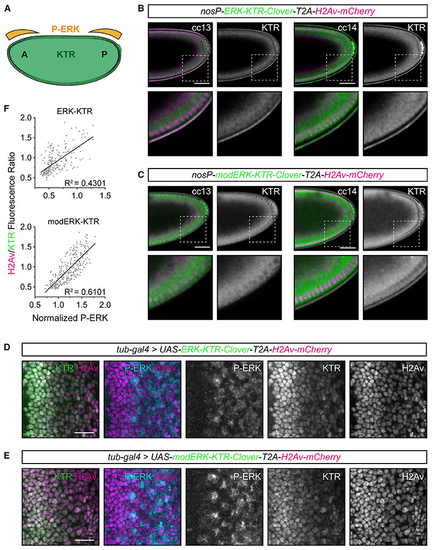
Improved Erk activity reporting with modERK-KTR in (A) Schematic of anteroposterior Torso/ERK signaling during early (B and C) Representative images of the posterior half of transgenic (D and E) Representative images of eye imaginal discs ubiquitously expressing ERK-KTR (D) or modERK-KTR (E) constructs with a polycistronic H2Av-mCherry tag under the control of (F) Quantification of (D) and (E) comparing P-ERK levels with the read out of ERK-KTR (n = 222 cells from 3 discs) or modERK-KTR (n = 293 cells from 3 discs) constructs and fitted with a simple linear regression. See also |
Reprinted from Developmental Cell, 58(23), Wilcockson, S.G., Guglielmi, L., Araguas Rodriguez, P., Amoyel, M., Hill, C.S., An improved Erk biosensor detects oscillatory Erk dynamics driven by mitotic erasure during early development, 2802-2818.e5, Copyright (2023) with permission from Elsevier. Full text @ Dev. Cell