
Figure 3
- ID
- ZDB-FIG-231205-3
- Publication
- Wilcockson et al., 2023 - An improved Erk biosensor detects oscillatory Erk dynamics driven by mitotic erasure during early development
- Other Figures
- All Figure Page
- Back to All Figure Page
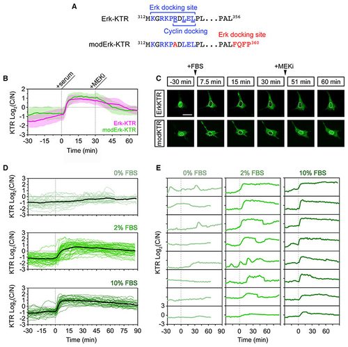
A modified Erk-KTR reports ERK activity in mouse fibroblasts (A) Amino acid sequences of the Erk-docking domain of Erk-KTR and modified Erk-KTR (modErk-KTR) highlighting the modifications (red) made to reduce off-target reporter activity, including the R>A substitution within the docking site and the additional FQFP Erk-docking site between the ELK fragment and the NLS. (B) Quantification of ERK activity in NIH-3T3 cells transfected with the Erk-KTR or modErk-KTR constructs (n = 58 cells each, mean ± SD). Cells were serum-starved overnight and ERK was induced by the addition of 10% FBS. ERK activity was inhibited after 30 min with 10 μM PD-0325901 (MEKi). (C) Representative images of reporter activity in (B). (D) Quantification of ERK activity in NIH-3T3 cells transfected with the modErk-KTR construct after overnight serum starvation, followed by the addition of different concentrations of FBS. Individual cell traces and the mean (black line) are shown for 0% FBS (n = 32 cells), 2% FBS (n = 57 cells), and 10% FBS (n = 30 cells). (E) Individual cell traces from (D). Scale bars, 20 μM. |
Reprinted from Developmental Cell, 58(23), Wilcockson, S.G., Guglielmi, L., Araguas Rodriguez, P., Amoyel, M., Hill, C.S., An improved Erk biosensor detects oscillatory Erk dynamics driven by mitotic erasure during early development, 2802-2818.e5, Copyright (2023) with permission from Elsevier. Full text @ Dev. Cell