|
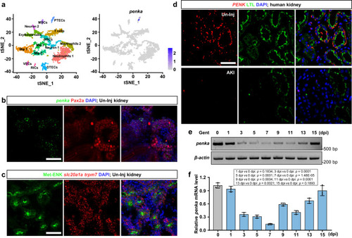
The expression patterns of zebrafish penka and human PENK in the kidneys. a scRNA-seq analysis revealed that penka was specifically expressed in zebrafish PTECs. t-SNE plots showing zebrafish kidney cell clusters and the expression of penka. VECs, vascular endothelial cells; DTECs, distal tubular epithelial cells; Mφ, macrophages; HSCs, hematopoietic stem cells; MSCs, mucin-secreting cells; and RICs, renal interstitial cells. b Confocal images showing double labeling of FISH-penka and anti-Pax2a in un-injured (Un-Inj) adult zebrafish kidney sections (n = 3). c Confocal images showing triple labeling of FISH-slc20a1a, FISH-trpm7, and anti-Met-ENK in Un-Inj adult zebrafish kidney sections. The Met-ENK signal co-localized with the signals of slc20a1a and trmp7, which are markers of PCT and PST, respectively (n = 3). d Confocal images of combined FISH-PENK and LTL staining in kidney sections of patients with AKI and patients with no detectable lesions (Un-Inj). Human PENK was expressed in PTs and downregulated after AKI (n = 3). e, f RT-PCR (e) and qRT-PCR (f) analyses of penka in zebrafish kidneys during Gent-induced AKI (n = 3). penka expression was decreased by 1 dpi and reached its lowest level at 7 dpi and returned to its un-injured level at 15 dpi. The data in (f) were analyzed by two-sided t-test and are presented as mean values ± SD. Scale bars in (b), (c), and (d), 50 μm. Source data are provided as a Source data file.
|