FIGURE

Fig. 2

ID
ZDB-FIG-231109-31
Publication
Liu et al., 2023 - Proenkephalin-A secreted by renal proximal tubules functions as a brake in kidney regeneration
Other Figures
All Figure Page
Back to All Figure Page
Fig. 2

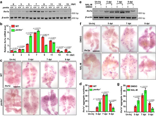
PENK-A deficiency accelerates kidney regeneration.

a RT-PCR analysis of lhx1a expression in WT and penka−/ zebrafish kidneys during Gent-induced AKI. b The band intensities in a are normalized to that of the loading control, β-actin, and the relative expression levels of lhx1a were quantified (n  =  3 biological replications per group). The data are presented as the fold change relative to the 0 dpi WT groups. c WISH analysis of lhx1a was performed in WT and penka−/− zebrafish kidneys without injury (Un-Inj) and at 5, 7, and 9 dpi. d Quantitation of lhx1a+ RPCAs (blue points) per kidney (n = 5) for each condition in (c). e, f RT-PCR (e) and WISH (f) analyses of lhx1a in Un-Inj, 5, 7, and 9 dpi kidneys after administration of NAL-M or DMSO (n = 3 in e, n = 4 in f). g The lhx1a+ RPCAs per kidney (n = 4) were quantified for each condition in (f). The data in (b), (d), and (g) were analyzed by two-sided t-test and are presented as mean values  ±  SD. Scale bars in (c) and (f), 600 μm. Source data are provided as a Source data file.

Expression Data

Expression Detail
Antibody Labeling
Phenotype Data

Phenotype Detail
Acknowledgments
This image is the copyrighted work of the attributed author or publisher, and ZFIN has permission only to display this image to its users. Additional permissions should be obtained from the applicable author or publisher of the image. Full text @ Nat. Commun.