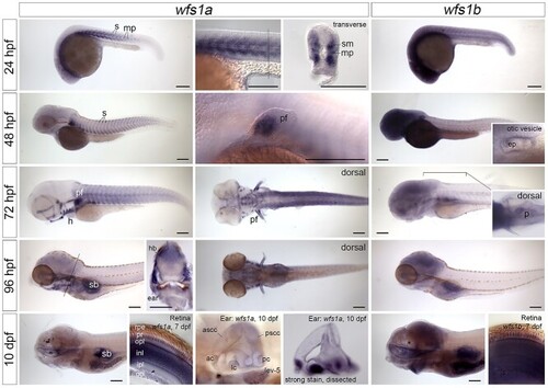
Expression of wfs1a and wfs1b mRNA in the zebrafish embryo, larva and juvenile. In situ hybridizations showing the expression patterns of wfs1a (left-hand and middle columns) and wfs1b (right-hand column) between 24 hpf and 10 dpf. All images are lateral views with anterior to the left, except for the inset showing expression of wfs1b in the pancreas (dorsal view, anterior to the left) and the sections through the retina. The position of the transverse hand-cut sections through the trunk at 24 and 96 hpf (dorsal to the top) is marked by a thin line in the preceding image. The eyes have been removed from the 10 dpf embryos, to show expression in the mouth and brain. The hand-cut thick sections through the retina, and the second image of the ear at 10 dpf, were taken from samples stained using an alternative protocol that resulted in stronger staining intensity (see Materials and Methods). Expression of wfs1a in the retina at 7 dpf is strong in the photoreceptors, outer plexiform and inner nuclear layers but is weaker or missing from the inner plexiform and ganglion cell layers (GCLs) and is very weak or absent from displaced amacrine cells (arrowhead). Expression of wfs1b in the retina at 7 dpf appears ubiquitous. Expression in the inner ear appears ubiquitous for both genes and may include some trapping. All images are bright-field images apart from the images of the trunk at 24 hpf, pectoral fin at 48 hpf and retinae at 7 dpf, which were taken with DIC optics. Abbreviations: ac, anterior crista; ascc, anterior semicircular canal; ep, epithelial projections in the otic vesicle; gcl, ganglion cell layer; h, heart; hb, hindbrain; inl, inner nuclear layer; ipl, inner plexiform layer; lc, lateral crista; lev-5, levator arcus branchialis 5; mp, muscle pioneer cells; opl, outer plexiform layer; p, pancreas; pc, posterior crista; pf, pectoral fin; pr, photoreceptors; pscc, posterior semicircular canal; rpe, retinal pigmented epithelium; s, somites; sb, swimbladder; sm, slow-twitch muscle fibers. Scale bars, 200 μm in all images.
|
