FIGURE

FIGURE 3

ID
ZDB-FIG-211111-28
Publication
Mayeur et al., 2021 - When Bigger Is Better: 3D RNA Profiling of the Developing Head in the Catshark Scyliorhinus canicula
Other Figures
All Figure Page
Back to All Figure Page
FIGURE 3

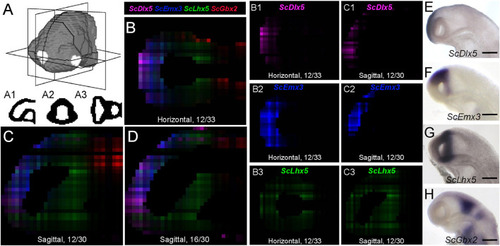
Digital profiles reproduce regional patterns along AP axis. (A) Scheme representing the digitized model of stage 17 catshark head with sagittal, transverse and horizontal digital section planes shown as white rectangles. (A1,A2,A3) show examples of digitized sections along these planes. (B–D) Digital horizontal (B) and sagittal (C,D) sections showing merged profiles of ScDlx5 (magenta), ScEmx3 (blue), ScLhx5 (green), and ScGbx2 (red). Sections are numbered from dorsal to ventral in panel (B) and from left to right in panels (C,D). (B1–3,C1–3) Digital sections showing the individual profiles, respectively, merged in panels (B,C) for ScDlx5(B1,C1), ScEmx3(B2,C2), and ScLhx5(B3,C3). (E–H) Left lateral views of stage 17 catshark embryonic heads after ISH with probes for ScDlx5(E), ScEmx3(F), ScLhx5(G), and ScGbx2(H). Scale bars = 200 μm.

Expression Data

Expression Detail
Antibody Labeling
Phenotype Data

Phenotype Detail
Acknowledgments
This image is the copyrighted work of the attributed author or publisher, and ZFIN has permission only to display this image to its users. Additional permissions should be obtained from the applicable author or publisher of the image. Full text @ Front Cell Dev Biol