FIGURE

Figure 1

ID
ZDB-FIG-210708-57
Publication
Salam et al., 2021 - Identification of a novel interaction of FUS and syntaphilin may explain synaptic and mitochondrial abnormalities caused by ALS mutations
Other Figures
All Figure Page
Back to All Figure Page
Figure 1

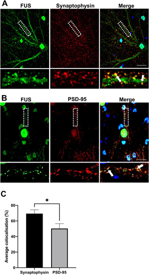
Subcellular localisation of FUS in primary cortical neurons. Immunofluorescent staining of DIV21 rat primary cortical neurons. (A) Representative confocal images of FUS (red) and presynaptic marker synaptophysin (SYN) (green) FUS was found to localise with synaptophysin puncta along neurites. (B) Representative confocal images of FUS (green) and the post synaptic marker PSD95 (Red). FUS was found to localise with PSD5 puncta along neurites. Selected regions of interest for A and B have been shown as magnifications as single and merged channels below the images of the respective neurons with white arrows indicating colocalisation. Nuclei are counterstained blue with DAPI. (C) Quantification of the subcellular localisation showed that FUS preferentially localised to the pre-synapse in these neurons (three neurites from nine different neurons from three independent experiments were analysed). *P < 0.05, unpaired students T-test. Scale bar = 20 μm.

Expression Data

Expression Detail
Antibody Labeling
Phenotype Data

Phenotype Detail
Acknowledgments
This image is the copyrighted work of the attributed author or publisher, and ZFIN has permission only to display this image to its users. Additional permissions should be obtained from the applicable author or publisher of the image. Full text @ Sci. Rep.