FIGURE

Figure 6

ID
ZDB-FIG-201003-72
Publication
Rima et al., 2020 - Dynamic regulation of the cholinergic system in the spinal central nervous system
Other Figures
All Figure Page
Back to All Figure Page
Figure 6

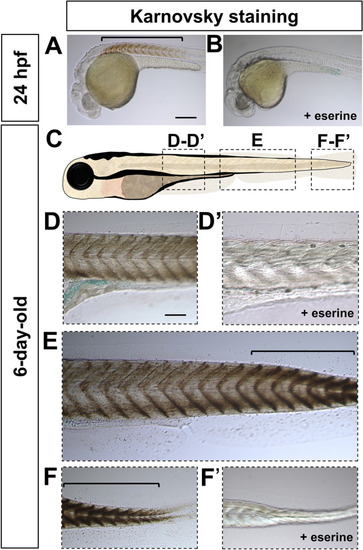
Spatiotemporal Acetylcholinesterase enzymatic activity pattern in the spinal cord during early development. (A, B) Lateral view of 24 hpf embryos processed with Karnovsky staining showing Acetylcholinesterase enzymatic activity (bracket) in the absence (A), but not in presence (B) of eserine, an AChE inhibitor. (C) Schematic representation of the imaged areas in (DF′). Lateral view of 6-day-old larvae showing a rostro-caudal gradient of AChE activity revealed by Karnovsky staining (DF′). (D, D′) Anterior trunk in the absence (D) or presence (D′) of eserine. (E) Mid trunk in the absence of eserine. (F, F′) Tip of the tail in the absence (F) or presence (F′) of eserine. Brackets indicate high levels of Karnovsky staining in (E, F). Magnification is the same for all images in (A, B) and (DF′). Scale bar: 200 μm.

Expression Data

Expression Detail
Antibody Labeling
Phenotype Data

Phenotype Detail
Acknowledgments
This image is the copyrighted work of the attributed author or publisher, and ZFIN has permission only to display this image to its users. Additional permissions should be obtained from the applicable author or publisher of the image. Full text @ Sci. Rep.