FIGURE

Figure 5

ID
ZDB-FIG-201003-71
Publication
Rima et al., 2020 - Dynamic regulation of the cholinergic system in the spinal central nervous system
Other Figures
All Figure Page
Back to All Figure Page
Figure 5

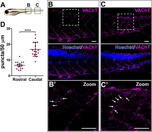
Decreased VAChT expression along the rostral myosepta of 6-day-old larva. (A) Schematic representation of the imaged areas in (B, C). (B, C) Confocal image of a lateral view of larva showing a rostro-caudal gradient of VAChT puncta in MN axons projecting along the dorsal myosepta. Nuclei are labeled by Hoechst (blue). (B′, C′) Zoomed in images of the boxed areas in (B, C). White arrows point toward VAChT positive signals along the myosepta. Scale bars: 50 μm. (D) Quantification of VAChT puncta in the dorsal myosepta. N = 4 larvae (15 rostral axons, 17 caudal axons). 2-tailed non-paired Student’s t test; ****p < 0.0001.

Expression Data
Antibody:
Fish:
Anatomical Term:
Stage: Day 6

Expression Detail
Antibody Labeling
Phenotype Data

Phenotype Detail
Acknowledgments
This image is the copyrighted work of the attributed author or publisher, and ZFIN has permission only to display this image to its users. Additional permissions should be obtained from the applicable author or publisher of the image. Full text @ Sci. Rep.