
Fig. S3
- ID
- ZDB-FIG-200810-7
- Publication
- Asokan et al., 2020 - Long-term in vivo imaging reveals tumor-specific dissemination and captures host tumor interaction in zebrafish xenografts
- Other Figures
- All Figure Page
- Back to All Figure Page
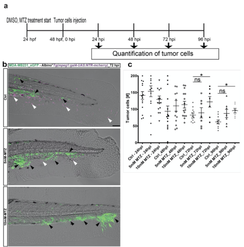
Ablation of macrophages revealed improved tumor cell survival. (a) Scheme depicting the experimental design: mCherry positive embryos were treated with either DMSO (for control) or MTZ (concentration: 5mM and 10mM) from 24 hpf. Every day, the embryo medium was changed with fresh DMSO and MTZ solution. 10 mM MTZ treatment resulted in 90% ablation of macrophages. Tumor cells were injected at 2 dpf and followed until 96 hpi. Every day embryos were analysed for tumor cell survival using confocal microscopy. (b) Representative image of GFP-labeled breast metastatic cells (BMC) (MDA-MB231) xenografted in eZXM expressing mCherry-labeled (magenta) macrophages at 72 hpi. In the top, control panel (ctrl), macrophages (magenta - white arrowhead) was observed in great numbers along with tumor cells (green – black arrowhead). 5mM-MTZ treatment showed hardly 2-3 macrophages (magenta - white arrowhead) were found and tumor cell number were comparatively higher to top control panel. 10mM-MTZ treatment in the bottom panel showed improved tumor cell numbers and almost no macrophages. Scale bar 100 μm. (b) Quantification of the tumor cells (BMC+) over time. At 72 hpi, a significant increase in the tumor cell survival was observed in the 10mM MTZ treated group compared to the control DMSO group. Plot represented means ± sem. Statistical analyses: one-way ANOVA followed by Dunnett’s test for multiple comparisons. Multiple comparisons: Ctrl_72hpi vs. 5mM MTZ_72hpi (P = 0.8699); Ctrl_72hpi vs. 10mM MTZ_72hpi (P = 0.0335); Ctrl_96hpi vs. 5mM MTZ_96hpi (P = 0.0667); Ctrl_72hpi vs. 10mM MTZ_72hpi (P = 0.0136). |