
Fig. S6
- ID
- ZDB-FIG-200810-10
- Publication
- Asokan et al., 2020 - Long-term in vivo imaging reveals tumor-specific dissemination and captures host tumor interaction in zebrafish xenografts
- Other Figures
- All Figure Page
- Back to All Figure Page
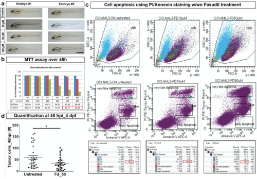
In vitro and in vivo effect of Fasudil. (a) Representative image of two embryo with different concentrations of Fasudil treatment to assess the phenotypic effect. Until 3 days post treatment (dpt), embryos did not show any phenotypic abnormality due to Fasudil treatments in comparison to control untreated embryos. Scale bar: 50 μm. dpt - days post treatment. (b) In in vitro culture, leukemic cells treated with Fasudil for 48 h showed a reduction of 80 % in their metabolic activity at 50 μM concentration using MTT assay (indicated by the values in red box). (Error bars are not indicated as experiment was performed only twice, a representative plot is shown here). (c) PI/Annexin staining on leukemic cells treated with Fasudil in in vitro for 24 h showed cells were apoptotic in comparison to control (left) as indicated by the green arrowhead in the second row of panel. Around 50 % of cells were only remaining after 50 μM Fasudil treatment as indicated by the red box in third row (right most bottom panel). (d) Quantification of tumor cells at 48 hpi in vivo in the zebrafish embryo showed a decrease in tumor cell number in 50 μM Fasudil- treated embryos [N=36 embryos]. Plots represent means ± sem. Statistical analyses: two-tailed Mann-Whitney’s U-test |