
Figure 6-S1
- ID
- ZDB-FIG-200406-187
- Publication
- Guillon et al., 2020 - Fibronectin is a smart adhesive that both influences and responds to the mechanics of early spinal column development
- Other Figures
- All Figure Page
- Back to All Figure Page
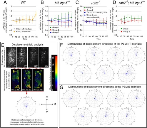
Medial-lateral ECM remodeling along the PSM|NT interface.(A–D) Quantification of the anterior-posterior length of the photoconverted FN matrix spots over time in wild-type PSM|NT and PSM|E interfaces (A), and in the PSM|NT interfaces in MZ itgα5-/-(B), cdh2-/- (C), and cdh2-/-; MZ itgα5-/- (D). In all plots, dots represent means and error bars represent SD. Sample sizes are as indicated in Figure 6. (E) Displacement field analysis of the photoconverted spots by particle image velocimetry (PIV). A displacement fields is constructed using a PIV plugin in ImageJ (see Materials and methods) using two successive time-frames. A frame-to-frame correlation of signals was established, and all vectors were plotted in a rose plot. A = anterior P=posterior M=medial L=lateral. (F–G). Distribution of displacement directions of the photoconverted regions at the PSM|NT (F) and at the PSM|E (G) interfaces in wild-type embryos. Each rose plot represents the data for one photoconverted region obtained as described in (E). In F, there is a bias toward anterior-lateral direction in six out of seven samples while there is no consistent directional bias in G. |