
Figure 3-S2
- ID
- ZDB-FIG-200406-181
- Publication
- Guillon et al., 2020 - Fibronectin is a smart adhesive that both influences and responds to the mechanics of early spinal column development
- Other Figures
- All Figure Page
- Back to All Figure Page
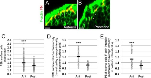
Epithelialization of PSM surface cells and increases in F-actin intensity in PSM cells as the PSM matures from posterior to anterior.(A, B) Transverse sections taken in the anterior PSM (A, 0–100 μm away from last somite boundary) or posterior PSM (B, 200–300 μm away from last somite boundary) on 12–14 s stage embryos co-stained for Fibronectin (FN, red) and F-actin (green). Asterisks denote PSM surface cells. Scale bars = 10 μm. (C) Quantification of PSM surface cell aspect ratio in the anterior and posterior portions of the PSM. Sample size: n = 93 cells (anterior PSM) and 92 cells (posterior PSM) from three embryos. Anterior vs posterior, p=7.46e-5. (D–E) Quantification of the mean F-actin intensity of the PSM surface cells region (D) or PSM internal cells region (E) in the anterior and posterior PSM from three embryos. Each dot represents a transverse section for which the mean F-actin signal within the surface cells or internal cells was measured and normalized by the average intensity of the signal in the posterior sections of the same embryo. Sample size: n = 24 sections (anterior PSM) and 25 sections (posterior PSM). ***p<0.0005 using a T-test. (D) Anterior vs posterior, p=5.62e-7. (E) (D) Anterior vs posterior, p=7.7e-4. |