FIGURE

Fig. S2

ID
ZDB-FIG-190604-57
Publication
Lan et al., 2019 - TETs Regulate Proepicardial Cell Migration through Extracellular Matrix Organization during Zebrafish Cardiogenesis
Other Figures
All Figure Page
Back to All Figure Page
Fig. S2

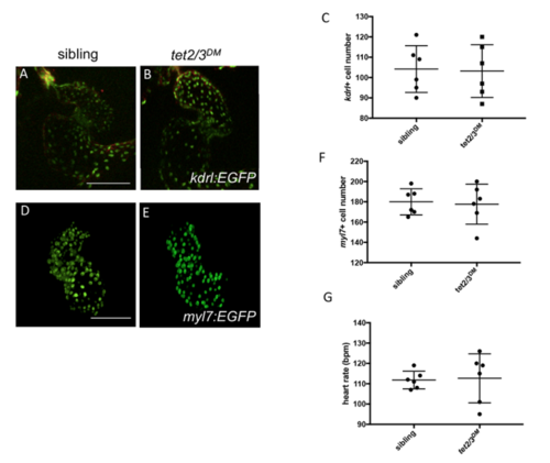
Normal Myocardium and Endocardium in 2-dpf tet2/3DM Larvae. Related to Figure 1.
(A and B) GFP labeled endocardium in 2-dpf larvae carrying the Tg(kdrl:EGFP-NLS) transgene.
(C) Graph indicates the number of endocardial cells in 48-hpf sibling and tet2/3DM larvae. Data are presented as the mean ± SD.
(D and E) GFP labeled myocardium in 2-dpf larvae carrying the Tg(myl7:EGFP) transgene.
(F) Graph indicates the number of myocardial cells in 48-hpf sibling and tet2/3DM larvae. Data are presented as the mean ± SD.
(G) Graph indicates heart rate in 48-hpf sibling and tet2/3DM larvae. Heart rate was measured in beats per minute. Data are presented as the mean ± SD.
Scale bars: 100 μm.

Expression Data

Expression Detail
Antibody Labeling
Phenotype Data

Phenotype Detail
Acknowledgments
This image is the copyrighted work of the attributed author or publisher, and ZFIN has permission only to display this image to its users. Additional permissions should be obtained from the applicable author or publisher of the image. Full text @ Cell Rep.