
Fig. 6
- ID
- ZDB-FIG-190604-55
- Publication
- Lan et al., 2019 - TETs Regulate Proepicardial Cell Migration through Extracellular Matrix Organization during Zebrafish Cardiogenesis
- Other Figures
- All Figure Page
- Back to All Figure Page
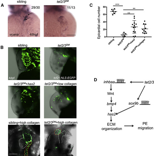
Tet2/3 Regulate PE Migration through Extracellular Matrix Organization (A) WISH for ECM constituent gene vcana at 48 hpf. Black arrows indicate AVC-specific expression of vcana in sibling, but not tet2/3DM heart. (B) GFP-labeled PE and epicardium in 4-dpf larvae carrying the Tg(tcf21:NLS-EGFP) transgene. Sibling, tet2/3DM, and tet2/3DMinjected with has2 mRNA and tet2/3DM injected with low concentration (0.5 mg/mL) collagen larvae were shown in lateral views. Sibling and tet2/3DM injected with high concentration (8 mg/mL) collagen larvae were shown in ventral views to represent collagen aggregate and heart clearly. The heart is outlined with white dashed line. The collagen aggregate is outlined with red dashed line. (C) Number of epicardial cells on the heart of 4-dpf sibling, tet2/3DM, and tet2/3DM injected with sox9b mRNA or tet2/3DMinjected with low concentration (0.5 mg/mL) collagen larvae carrying the Tg(tcf21:NLS-EGFP) transgene. Numbers data are presented as the mean ± SD derived from 3 independent biological replicates. (D) Working model shows Tet2/3-dependent demethylation regulates the expression of inhbaa and sox9b, which subsequently regulate AVC ECM organization and PE migration. Scale bars: 50 μm. The significance is indicated as ∗p < 0.05; ∗∗p < 0.01; ∗∗∗p < 0.001 |
| Gene: | |
|---|---|
| Fish: | |
| Anatomical Term: | |
| Stage: | Long-pec |
| Fish: | |
|---|---|
| Observed In: | |
| Stage: | Long-pec |