
Fig. S1
- ID
- ZDB-FIG-190604-56
- Publication
- Lan et al., 2019 - TETs Regulate Proepicardial Cell Migration through Extracellular Matrix Organization during Zebrafish Cardiogenesis
- Other Figures
- All Figure Page
- Back to All Figure Page
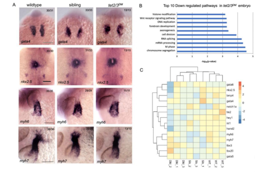
tet2/3DM Larvae Show Neuronal but not Cardiac Defects at 28 hpf. Related to Figure 1. (A) Markers of cardiac progenitors are similarly expressed in wildtype, sibling and tet2/3DM larvae. WISH forgata4 was performed at 18 hpf, nkx2.5 was performed at 22-hpf, atrial myosin marker myh6 and myosin marker myh7 were performed at 28-hpf. Scale bar: 100 μm. DM (C) Heatmap of RNA sequencing data illustrating similar transcriptional expression of cardiac genes in tet2/3(DM) compared with wildtype (WT) or sibling (Sib) larvae. DM |